måndag 10 augusti 2009

Chakras and the colors II. Love and pain.

Light in our biology, in our head.
Are there anything in the biology that possibly could explain this light to us? The light gives euphoria, joy, love, strength, wisdom. It is a psychedelic drug. In biology we know of endorphins giving pleasure, phenethylamine PEA is addicting in love, sex stimulates the release of vasopressin and oxytocin, DMT makes you see God, and so on.

So, I read an interesting paper. Coordination involves a subtle kind of ballet in the brain, and like dancers, cortical areas are capable of coming together as an ensemble (integration) while still exhibiting a tendency to do their own thing (segregation). What? Quantum biology in working?
Because of the fourth dimension afforded by this colorimetric method, it is possible to observe and interpret oscillatory activity of the entire brain as it evolves in time, millisecond by millisecond, and show how to tell apart real and false episodes of synchronization. For the first time, true episodes of brain coordination can be spotted directly in EEG records and carefully analyzed.

The brain areas active in love are different from the areas activated in other emotional states, such as fear and anger. Parts of the brain that are in love include the one responsible for gut feelings, and the ones which generate the euphoria induced by drugs such as cocaine. So the brains deeply in love do not look like those experiencing strong emotions, but instead like those of people snorting coke. Love, in other words, uses the neural mechanisms that are activated during the process of addiction.

Strong social bonds depends on the receptors for vasopressin and oxytocin. Evolution acts on the distribution of these receptors to generate social behavior. The more receptors located in regions associated with reward, the more rewarding social interactions become. There are variations between individuals. But once romantic love begins, it is one of the strongest drives on Earth. This power is enough to warp judgment in otherwise sensible people. Althruism is also a social bond, or a freeing from the singular I.

Some suggest this mental state might share neurochemical characteristics with the manic phase of manic depression. Means love can be "treated" pharmacologically (serotonin-inhibitors)? It might even to be possible to create a more sociable human? And what about a more loving one? A few people even think that “paradise-engineering” is possible?

The experience of romantic attraction activates those sites in the brain with a high concentration of receptors for dopamine, closely tied to states of euphoria, craving and addiction. "The brain activity pattern is markedly different from when they looked at a picture of a close friend," reported neurobiologists Andreas Bartels and Semir Zeki. They linked high levels of dopamine and norepinephrine to heightened attention and short-term memory, hyperactivity, sleeplessness and goal-oriented behavior. Bartels and Zeki compared brain scans taken from people in different emotional states, including sexual arousal, feelings of happiness and cocaine-induced euphoria. The pattern for romantic love was unique. But there was some overlap with and close proximity to other positive states. "It's this combination of friendship, affection and lust, that makes it so powerful."

And where in the brain is this light?
Let us look at some conditions. There are many parameters that make up the control levels. Not one frequence or amplitude, nor one molecule, but a parameter space of multidimensional character (a 4-D brain as Matti Pitkänen says, with time as one parameter?).
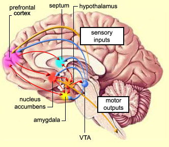

The brain’s reward circuit are located along the medial forebrain bundle (MFB). The ventral tegmental area (VTA) and the nucleus accumbens are the two major centres in this circuit, but it also includes several others, such as the septum, the amygdala, the prefrontal cortex, and certain parts of the thalamus. All of these centres are interconnected and innervate the hypothalamus (red arrows), informing it of the presence of rewards.

1. Love and addiction areas. The vasopressin and oxytocin receptors were equally distributed in the brains of male and female voles, according to Dr. Young, coupled with the close proximity of the nucleus accumbens and ventral pallidum - two regions with heavily interconnected structures - suggest that a common neural circuit in male and female voles regulates pair bond formation. The dopamine system of the nucleus accumbens produces the rewarding and sometimes addictive effects of sex, food and drugs of abuse. The receptors are closely located to the nerve fibers that release oxytocin and vasopressin.

Using fMRI to view the brains of easily orgasmic women as they climaxed, either with visual stimuli or by self-stimulation, Whipple found that the body's pain-killing center in the midbrain is activated during peak arousal. Signals from this part of the brain instruct the body to release endorphins and corticosteroids, and is also coupled to anxiety.

2. Feel good, feel bad. The pleasure system includes the septal area and part of the almond-shaped amygdala; the other half of the amygdala, the hippocampus, the thalamus, and the tegmentum (in the midbrain) constitute the punishment system (rage, anxiety, pain). The flip side of joy is pain. Whenever a mental patient flew into a violent rage or turned into a catatonic zombie, the EEG was almost certain to display the telltale sawtooth pattern. If the patient got well, the spike disappeared. ...

3. Schizophrenia. "The primary symptom of schizophrenia isn't hallucinations or delusions," says Heath. "It's a defect in the pleasure response. Schizophrenics (negative symptoms?) have a predominance of painful emotions. They function in an almost continuous state of fear or rage, fight or flight, because they don't have the pleasure to neutralize it." ... It turned out that electrical stimulation of the pleasure center automatically turned off the punishment system. The cerebellum, Heath learned, is a better entryway to the brain's emotional circuitry. Stimulating a precise half-inch of its cauliflowerlike surface automatically fires the pleasure area and inhibits the rage centers.

[From a patient story:] "There -- see the big delta wave appearing in the septal region. Sure enough, large, languorous waves are now coming from the lead to the septal electrode. There's almost an exact correlation. When he gets a rush of good feeling, the record shows large-amplitude waves in the pleasure system."" ...

Along with depth electrodes, Heath's team would often surgically implant a sort of tube, a canula, through which they could deliver precise amounts of a chemical directly into the brain. Oriental sacred texts (and Aldous Huxley's Brave New World) mention a legendary bliss drug called "soma", the food of the Himalayan gods. The real life version might be acetylcholine. When acetylcholine was injected into a patient's septal area, "vigorous activity" showed up on the septal EEG, and the patient usually reported intense pleasure - including multiple sexual orgasms lasting as long as thirty minutes. An ominous-looking scrawl on the EEG looks almost like the spoke-and-dome pattern of epileptic seizure. It's a very explosive activity. (Afrodisiaca is often acetylcholinic).

4. Psychosis. Nucleus accumbens is the key structure of the brain responsible for reward, motivation and addiction. Addiction, motivation and impulsivity has much in common, also learning. Amphetamine-like drugs (which increase dopamine levels by blocking its re-uptake) are the main drug treatment for Attention Deficit/Hyperactivity disorder of which impulsivity is a component.

Psychosis refers to a subset of the symptoms associated with schizophrenia - symptoms which include hallucinations and delusional thoughts. It is rather striking that although there is no cure for all of the symptoms of schizophrenia, there is a single pharmacological treatment for the psychotic symptoms - blockade of dopamine receptors. Nobody knows why an excess of dopamine might lead to delusional thoughts or hallucinations, but it seems rather remarkable that the control of a single type of molecule in the brain is capable of influencing a person's thought processes in such a selective fashion.

Dopamine is a neuromodulator released by dopaminergic cells that originate in the midbrain (Ventral Tegmental Area (VTA) and Substantia Nigra) and which project to a number of brain regions including the Prefrontal Cortex (PFC), the amygdala, the hippocampus and the striatum (Caudate nucleus). All of these brain regions have been implicated in schizophrenia, although perhaps the most interesting region of all is the nucleus accumbens. The nucleus accumbens is connected to all of these other important regions, and is known to contain a large proportion of D2 receptors. The importance of the D2 receptor in particular is suggested by the striking correlation between the ability of an antipsychotic drug to block this receptor and the ability of that drug to mitigate the symptoms of psychosis. What does the nucleus accumbens do in the brain? What does the D2 receptor do? When and why is dopamine released? What is the functional role of dopamine?

5. Migraine and epilepsy. In certain syndromes the two conditions epilepsy and migraine are associated, characterized by visual symptoms followed by a partial seizure and postictal migraine. The EEG reveals occipital spikes (visual?). The visual symptoms are often followed by seizures. After the seizure, approximately 25–40% of the patients develop migrainelike headaches with a migrainous aura.

Another epilepsy-syndrome is characterized by unilateral somatosensory or motor seizures and centrotemporal spikes. Clinical and electrographic features can shift from side to side. Speech arrest, pooling of saliva, and usually preservation of consciousness are also typical, although spread and generalization do occur. Epileptic seizures are predominantly multi-coloured with circular or spherical patterns as opposed to the predominantly black and white linear patterns of migraine. Also those of epileptic patients are predominantly centrally localized, whereas those of migraine patients are predominantly peripherally localized.

Attention-deficit hyperactivity disorder (ADHD) is also much more common in epileptics (20%) as compared to normal children (3-7%). Several mechanisms may account for the high prevalence, such as a common genetic propensity, noradrenergic system dysregulation, subclinical epileptiform discharges, or even seizures, antiepileptic drug effects. Children with attention-deficit hyperactivity disorder have a higher than normal rate of EEG abnormalities. The noise or the input level is much higher, and so the attention fades. Also memory can be halted.

The clinical diagnosis of visual seizures is easy if individual elements of duration, colour, shape, size, location, movement, speed of development and progress are identified. They are markedly different from visual aura of migraine, although they often trigger migrainous headache, probably by activating trigemino vascular or brain stem mechanisms.

6. Pain on/off. The trigeminothalamic tract and periaqueductal grey (PAG). Also indicates that the thalamocortical tract is an important component of migraine pathophysiology. The central sensitization observed in migraineurs is most probably related to an abnormal activity of the trigeminal sensory pathway. A possible dysfunction of the periaqueductal grey in migraineurs without aura, which could result in a lowering of the threshold for initiation of migraine attack through a lack of inhibition of the trigeminal sensory activation.

As a critical pain neuromodulating structure, the periaqueductal grey matter has been implicated with the activation of the nervous system in migraine. The ventrolateral periaqueductal grey is part of a descending paininhibiting system from the hypothalamus and frontal cortex projecting to the medullary and spinal dorsal horns. Periaqueductal grey matter activation inhibits contralateral trigeminovascular nociception. PAG was hyperactive during migraine. The exact neurotransmitters behind the aura are unknown, ev. NO. PAG is also involved in narcotic abuse.

The PAG has a large influence on the nociceptive pathways (pain treshold)(perception of pain = treshold value for pain) with extensive networks from thalamus, hypothalamus and autonomic nervous system. It is full of opiate receptors (endorphins and others) according to Pert, and is a control area for pain, and also for most of other peptides/receptors. Pain is a highly complex and with subjective experience that is not linearly related to the nociceptive input. It is well known that pain perception for patients and normal subjects can be modulated by psychological factors, such as attention, stress, and arousal. Activation in the periaqueductal gray was significantly increased when pain was inhibited. In many sensory modalities, afferent processing is dynamically modulated by attention and this modulation produces altered sensory experiences.
An increase in flow of the middle meningeal artery (MMA) also occured. Pert says that pain can be altered by expectanses, faith and it can even be altered into pleasure. What is the role of consciousness in this, asks Pert.

Trigeminus input to the brain has also a profound impact on the consciousness level according to Damasio. Pain is a conscious experience, an interpretation of the nociceptive input influenced by memories, emotional, pathological, genetic, and cognitive factors. Is consciousness dependent on perception (of pain = disturbance or noise?)? Or attention? PAG is also our first body map in the nervous system. And pain and serotonin is dependent on each other. Paininhibition means serotonin excreation with urine. Adrenaline and noradrenaline is the counterpart. Noradrenaline is happiness, says Pert. Is our consciousness dependent on a disturbance, or arousal? Sounds a simple question, but it isn't. These painmodulations can be initiated reflexively or by contextual manipulations of the pain experience including cognitive and emotional factors. This provides a necessary survival function since it allows the pain experience to be altered according to the situation. Plasticity, sensitization and other amplification processes might occur along the pain neuraxis for an individual and one can relate this to their specific pain experience or measure of pain. Individuals can gain voluntary control over activation in a specific brain region given appropriate training (biofeedback as ex.), that voluntary control (over behavior and cognition) over activation in the rostral anterior cingulate cortex (rACC), leads to control over pain perception, and that these effects are powerful enough to impact severe, chronic clinical pain. Pert also points out that breeding is a control of pain. Forced breeding make more peptides and of a different kind in the brain stem. They are then expressed in PAG.

7. Gender, developement, morphology, cognition and mood; the Dentate gyrus in hippocampus. In the late 1980s, the finding that the dentate gyrus contains more granule cells in the male than in the female of certain mouse strains provided the first indication that the dentate gyrus is a significant target for the effects of sex steroids during development. Gonadal hormones also play a crucial role in shaping the function and morphology of the adult brain. Besides reproduction-related processes, sex steroids participate in higher brain operations such as cognition and mood, in which the hippocampus is a critical mediator. Sex steroids modulate the function of dentate neurons under normal conditions. In addition, recent research suggests that hormone-induced cellular plasticity may play a larger role than previously thought, particularly in the dentate gyrus. Specifically, the regulation of dentate gyrus neurogenesis and synaptic remodeling by sex steroids, and gonadal hormones have neuroprotective potential. Gonadal hormones could also influence the dentate gyrus indirectly, by subcortical hormone-sensitive structures such as the cholinergic septohippocampal pleasure system.

Serotonin has also been linked to the effect of estradiol on dentate neurogenesis. Indeed, several studies indicate that estrogen influences the dentate serotonergic system, f.ex. estradiol treatment reduces serotonin gene expression in the dentate gyrus. That's perhaps the reason why menopause has been linked to kundalini-experience.

8. The secondary sexual behavior is also related to the anterior cingulate gyrus (in the roof of the limbic area), as a response of vasopressin and oxytocin receptors. Care for the offspring belongs here, as well as aggressive behavior (male). Large bilateral leisons abolish voluntary movement, but not awareness. "Nothing matters", and "I had nothing to say", said a patient. Smaller leisons gave severe depression and anxiety. Or loss of voluntary control of a hand (alien-hand syndrome), that has a will of its own. (Brain-wise by Churchland).


Selfcontrol over sexual arousal belongs here. Limbic areas can be controlled by free will. Autistic people have disturbances in cingulate gyrus (small cells, high density), hypothalamus, amygdala, mammillary bodies and cerebellum.

Motivation and decision-making belongs here too, orchestrated by serotonin and dopamine, norepinephrine and acetylcholine, as well as various hormones, estrogen and testosterone. Judgement and impulse control can fail due to too much estrogen or testosterone.

Control by suppressing emotions, feelings and inclinations is often believed to be rational (and virtue). But reason can never be a motive, or make decisions. Reason can't make free will, can't give aversion against something or a drawing to something. Reason can tell lies, storytellings and fantasies. Feelings is against or for, a common sense. Emotions is a good thing, but too much emotions and too much reason, virtue and moral can be fatal. As a rule strong passion can't be controlled, too much reason and you go astray.

9. The pineal gland is unique in its solitary status within the brain. All other brain sites are paired, meaning that they have left and right counterparts; for example, there are left and right frontal lobes and left and right temporal lobes. As the only unpaired organ deep within the brain, the pineal gland remained an anatomical curiosity.
The pineal gland of evolutionarily older animals, such as lizards and amphibians, is also called the "third" eye. Just like the two seeing eyes, the third eye possesses a lens, cornea, and retina. It is light-sensitive and helps regulate body temperature and skin coloration—two basic survival functions intimately related to environmental light (and emotions).

What have transcerebral, weak (1 microT) complex magnetic fields and mystical experiences in common? Are they generated by field-induced dimethyltryptamine (DMT) release from the pineal organ? Structurally, DMT is analogous to the neurotransmitter serotonin and other psychedelic tryptamines. Some believe it plays a role in mediating the visual effects of natural dreaming, and also near-death experiences, religious visions and other mystical states. In addition to being involved in altered states of consciousness, endogenous DMT may be involved in the creation of normal waking states of consciousness. Dimethyltryptamine dose dependently elevated blood pressure (tonus), heart rate, pupil diameter, and rectal temperature, in addition to elevating blood concentrations of beta-endorphin, corticotropin, cortisol, and prolactin. Growth hormone blood levels rose equally in response to all doses of DMT, and melatonin levels were unaffected. DMT and other endogenous hallucinogens mediate their neurological abilities by acting as neurotransmitters at a sub class of the trace amine receptors?

Decreased sensitivity to the serotonergic hallucinogens psilocybin and LSD is induced by drugs with effects on serotonergic neurotransmission, allopurinol and fluoxetine?

Hallucinogens' effects in humans are mediated by serotonergic receptors?In this way waking consciousness can be thought of as a controlled psychedelic experience, or stress induced by environment and giving rise to muscle work? It is when the control of these systems becomes loosened and their behavior no longer correlates with the external world that the altered states arise. A theory was also that a massive release of DMT from the pineal gland prior to death or near death was the cause of the near death experience (NDE) phenomenon. Several test subjects reported NDE-like audio or visual hallucinations. The explanation for this experience was the possible lack of panic/pain.

Attention and concentration is also an effect of serotonin, as well as violence and suicide can be that. In fact serotonin is very central to our human being charachters. This is something I have called "the serotonine wheel" for my own purposes. Are there also a "dopamine wheel", an endorphine wheel" etc.?

One of the most obvious physiologic manifestations of seasonality is the phenomenon of seasonal breeding. The pineal gland hormone, melatonin,mediates the antireproductive effects of decreased light exposure during winter months. Annual rhythms in humans have also been established, including, for example, seasonal variations in suicides, general mortality, a need for electroconvulsive therapy and the incidence of episodes of depression and mania. Melatonin is made from serotonin.

Pineal gland is very sensitive to magnetism. How is it with normal visual perception and magnetism? Pineal gland can easily be calcified with "brain sand" (fluor) and so can be hypofunctional.

Was Descartes right after all?
We shall continue with the God-concept. See also http://zone-reflex.blogspot.com/2009/07/chakras-and-colors-i.html

References
See also links.
Dynamical Theory and Novel 4D Colorimetric Method Reveal the Essential Modus Operandi of the Intact Living Brain--Study shows how areas in the brain integrate and segregate at the same time. Newswise. See also Tognoli, E., Kelso, J.A.S., Brain coordination dynamics: True and false faces of phase synchrony and metastability. Prog. Neurobiol. (2008), doi:10.1016/j.pneurobio.2008.09.014

Sheena K Aurora. Pathophysiology of Migraine. NeuroScience 2007 - Supplement to EU/US Neurological Disease 2007 Issue 1 (BTG). http://www.touchneurology.com/files/article_pdfs/neuro_7463

Knight Y.E., Goadsby P.J. The periaqueductal grey matter modulates trigeminovascular input: a role in migraine? Neuroscience, Volume 106, Number 4, 31 October 2001, 793-800(8).

Afridi and Goadsby. New onset migraine with a brain stem cavernous angioma
J Neurol Neurosurg Psychiatry.2003; 74: 680-681 http://brain.oxfordjournals.org/cgi/content/abstract/125/6/1392 - The selective low efficacy adenosine A1 receptor agonist, GR190178 (30–1000 µg/kg i.v.), also inhibited SSS-evoked neuronal activity in a dose-dependent fashion. In this model of trigeminovascular nociception, adenosine A1 receptor activation leads to neuronal inhibition without concomitant vasoconstriction,

C P Panayiotopoulos: Elementary visual hallucinations in migraine and epilepsy. J Neurol Neurosurg Psychiatry. 1994 November; 57(11): 1371–1374. http://www.pubmedcentral.nih.gov/articlerender.fcgi?artid=1073189

Michael A. Rogawski 2008: Common Pathophysiologic Mechanisms in Migraine and Epilepsy. Arch Neurol. 2008;65(6):709-714. http://archneur.ama-assn.org/cgi/content/abstract/65/6/709

http://www.ncbi.nlm.nih.gov/pubmed/18603235?ordinalpos=1&itool=EntrezSystem2.PEntrez.Pubmed.Pubmed_ResultsPanel.Pubmed_DiscoveryPanel.Pubmed_Discovery_RA&linkpos=5&log$=relatedreviews&logdbfrom=pubmed

Tibor Hajszan, Teresa A Milner,and Csaba Leranth, Sex Steroids and the Dentate Gyrus. Prog Brain Res. 2007; 163C: 399–816. http://www.pubmedcentral.nih.gov/articlerender.fcgi?artid=1964752.

Sculthorpe L, Persinger MA. Does phase-modulation of applied 40-Hz transcerebral magnetic fields affect subjective experiences and hypnotic induction? http://www.ncbi.nlm.nih.gov/pubmed/15002842?dopt=Abstract

Patricia Smith Churchland, 2002: Brain-Wise: Studies in Neurophilosophy. Cambridge. MIT Press.

lördag 1 augusti 2009

The Wickman thing

I just met a "brandnew" cousin, 54 years old. Just one year older than me. I have met her once before, in the sixties, when I was 6-7 years old. I remember that just vaguely. The Wickman family has many relatives "over there". In fact half the family? Some went long ago, and their children and grand children is now looking back, trying to find their roots. Monica too had her daughter with her. She was in the same age as my son.

Mostly Monica has just been a name on a paper, just as all the other cousins I have "over there". Monica. I remember I have heard my mam and dad speak of her and her three brothers too. She has lost contact with one of her brothers, the other ones she hardly ever meet.


Monica lived her childhood i Vesterås in Sweden. Her daddy was my uncle. I don't remember him at all, although I must have met him as a child. Her daddy was a person who moved a lot. He didn't think long for a decision, he just did it. He was quite stubborn too. (Guess from where I have got that one?) Nobody could tell him what to do. Monica told stories about my relatives and made them more living to me. And so I got to learn something about myself too, perhaps not so good things, but things I have wondered about. Why do I react at things that happens in the way I do? My ability to make fast decisions sometimes. You may ponder long, then something happens, and suddenly you just know. And you have to follow your idea.

Her daddy was fast to make decisions. She told when she was thirteen and her brother was 9, her dad took them from home to Whiterocks in British Columbia, quite a bit, in fact as almost half the Finland, and he liked the place, so he decided that they should live there. He rented a room and left Monica and her brother there alone. He himself went home packing. He took his wife and two other sons with him to Whiterocks. Fortunately they also was pleased with their new homes. Monica said "But what, if he had an accident on his way home? Nobody else knew where we were". That was Gustav, my uncle. I know it too well. Quess how many stories my parents have told me about my childhood and my ability to always make stupid things. It was my speciality. Monica said it was "the Wickman thing. All Wickmen do stupid things, but they are also very brave and do hard work. But so hopelessly stubborn!"

But it came to a divorce after a few years for my uncle.

Her daddy never told where he should go, or how long he would stay. Monica told me that for some reasons:-) she was quite the same. There were many stories of that kind that evening. Of course without reason:-) They never made such things it you asked them. No, no, no. Never.

And I am also like that. Quess why?
Only afterwards I can laugh. I laugh a lot.

söndag 26 juli 2009

Chakras and the colors I

The color frequencies has much in common with the chakras of Eastern Wisdom. How can the chakras be interpreted? Are there any science pointing to their existence? Let us first look at the ancient teachings in short (very short, indeed).

"The Yellow Emperor" is a classicer in acupuncture, dating back to Emperor Fu Shi (2300 BC), who obtained the true Tao. The book has a true historical value, and can well be compared with the holy Bible. A sacred diagram on the body of a dragon horse was "revealed" to him, with fifty-five dots of “Yin” and “Yang” to represent the five elements. He then observed the Heaven and Earth, studied the conditions of the environment, habits and forms of animal and bird. From these observations, he started to formulate the eight trigrams (I Ching) for the purpose of helping people to realize the virtue of Heaven to recover the true nature of mankind. The lines are called lines of change, a process, a stribe to, a possibility, Yin, and its realization Yang, in cosmos.

(In Egypt Kheper and kheperu (plur.) were related to the process of spiritual growth, self-creation, and change. The sacred scarab beetle, Khepri, symbol of the Great mother, transformation, being and becoming. Deities for the Kheperu was Atum, the creation god I, and Isis, the female god over all. Priestess and priests of Isis actually becomes gods for a time in the rites of the House of Isis.)

The beginning, cultivation, reward and perseverance are kind of an evolution. The beginning, Quan, is creation. It represents “Tao” in the cosmos and the True Self in human. Truth, reward, indicates that all things and beings are at proper places and in harmony. The root of all substances and matters is that all beings following the everlasting Tao ("Tao of Heaven" is the endless regeneration of this cycle). Tao is change and movement.

The Monad Te (evolution) is non-reliable and chaotic Tai Chi, which are in between two forces: the Yang, negenthropy or expanding, growing energy that will become matter, and it will be bent backwards into a ball; and the Yin, enthropy or matter that will become energy, a line. Information is inside the monad and the monad will go through the evolutional process, in forming atoms, molecules and cells, organs, organisms, and all the different creatures. Every monad Te comes from the Tao potential and has therefore its own place in the harmony.

Every creature is between the forces of time, Yang, and space, Yin. Also in between heaven, the endless Kiä, and the earth, the bounded one, Kun. Earth is a line, because of the gravitation; the heaven is a ball because of the sun and the planets. So the human is concentrated in between the earth, the human and the heaven, in the focus of a triad, with the essence of Chi, the potential of heaven. Only when the human triad is empty he will experience his existence as a focus of these forces. The human being can rest in an empty house, Tandien, and control every movement and situation by taking energy, Yin, or giving energy, Yang. Yin and Yang is not fixed but relative.

Aura and chakra
We all have an aura surrounding our bodies. This aura is of different colours, different sizes and different qualities. It is as you really was different bodies, but you experience yourself as being one whole individual, a self or singular I. Without that aura nothing can exist. The auras regulate all functions in the body, all potential forces, and all the structures. The aura is a summary of the body itself and its functions and structures, and the outside world and its forces and energies. The chakras will recieve, change and regulate the energy flowing through. The chakras will also radiate out energy from the body into the aura and further out into the space.


With higher frequencies you have higher consciousness-levels. With higher energy amplitudes or pool you have a larger aura. . See the electromagnetic spectrum. (The behavior of EM radiation depends on its wavelength. Higher frequencies have shorter wavelengths, and lower frequencies have longer wavelengths. When EM radiation interacts with single atoms and molecules, its behavior depends on the amount of energy per quantum it carries. Spectroscopy can detect a much wider region of the EM spectrum than the visible range of 400 nm to 700 nm.)


Fig. Aura, an example, from Leadbeater, C.W., 1980: Man visible and invisible, 3rd ed. The Theosophical Publ. House. USA.

The chakras and energy centers in the body are seven (eight) greater and some smaller (out of body there are much more). The scripts mention as many as 88000 and the more meaningful chakras are 40. All the acupuncture points are small chakras. Keeps the different energy bodies in contact with each other. Energy vessels like “nadis” or “meridians”; s many as 350000 nadis is mentioned in historical scripts. Often it is not meaningful to discriminate that many. In Chinese scripts they usually elaborate with 12 primary meridians (organ) and about six other meridians (extraordinary), with other combinations of points. In India they have three prime nadis; sushumna, Ida and Pingala. Sushumna is inside our spine, in the cannel, Pingala is the sun´s energy, which is a glowing, shining force and begins in a petal in the root chakra at the right and stops at the right nose wing, sieraimen. Ida is cooling, calming down, and is energy from the moon. This starts to the left and ends to the left; the opposite to Pingala. They make a spiral movement around the viscerals and around sushumna. Ida and Pingala can take prana/Chi directly from the air and transport it inside, and when we breath out we get rid of poisons.

When Chi is manifesting itself it manifest through many different frequencies and consciousness-levels. Breathing is one of them. But in no way oxygen is equal to Chi.
Different kind of creatures is receptive for different kinds of energy. So animals have lower frequencies in their aura than human beings. This is seen as different colours.

The Caduceus was originally a healing tool employed in the temples of Atlantis and Egypt. It had been brought into this realm by the Illumined Master, Thoth. The wings on the Caduceus are symbolic of the liberation of consciousness from the warp and weave of dual systems, once it moves up the staff between the serpents and further, beyond their reach. The thyrsus was a device with the same meaning as the caduceus.

If we compare with an aura and chakra wheels. See the yellow spots in the hands and along the spine, together with a yellow head.


Let us compare to the colors.
Where one monochromatic color ends and another begins is a matter of debate as you will see in the table below.

Wavelength Ranges for Monochromatic Light (nm)
color   1 2 3 4
red   647-700 647-760 630-700 620-800
orange   585-647 585-647 590-630 590-620
yellow   575-585 575-585 570-590 560-590
green   491-575 491-575 500-570 480-560
blue   424-491 424-491 450-500 450-480
violet   400-424 380-424 400-450 400-450
1. CRC Handbook of Chemistry and Physics. 1966.
2. Hazel Rossotti. Color. Princeton University Press, 1983.
3. Edwin R. Jones. Physics 153 Class Notes. University of South Carolina, 1999.
4. Deane B. Judd. Goethe's Theory of Colors. MIT Press, 1970.


Compare with the chakras, violet is the crown.

"The secret of the golden flower"
In India the theory of the three elements in the Upanishad led to the theory of the three forces, and to the later theory of five elements. Also in Tibetian medicine they uses three humorals and five elements. In China, the theory of five elements coexisted early with the theory of two forces: Yin and Yang. Chi is composed of three immaterial parts.

The chakras are often pictured as lotus-flowers about 10 cm in diameter, with different amounts of petals. The petals are a symbol for nadis, meridians. From the flowers you have stalks going to your channel, sushumna, in your spine, the energy goes down-up. The chakras are always circulating like wheels. This is the force driving the energy into sushumna. If you change the direction of the circulation, energy will run out. The chakras circulate always in a special manner, different for male and female. Also different for different wheels (colors).

Different chakras have also different colours (see auras), different senses, different consciousness-levels. They can be quite different in different persons and in the same person from time to time. The size and colour regulate the quality and amount of energy flowing through.

Kundalini, Shiva and Shakti, Fire and Water, to climb the mountain
Kundalini (Sanskrit kund, “to burn”; kunda, “to coil or to spiral”) is a concentrated field of intelligent, cosmic invisible energy absolutely vital to life; beginning in the base of the spine when a man or woman begins to evolve as wisdom is earned. Kundalini has been described as liquid fire and liquid light. You may feel it when it pulates. The ultimate outcome of kundalini is the union of Will (shakti- kundalini), Knowledge {prana-kundalini) and Action (para- kundalini). When that light rise it is "the yellow flower". The light (yellow or white) burns inside your head. To get your kundalini to rise (yellow light along your spine) you must have enough input of energy (sexual, conscious, emotional, accidential or psychedelic drugs as mescaline, LSD). And the more easy flowing, the bigger it will grow, and so its consciousness.

Energy can also come in from outer world via nadis, meridians and acupoints. But in general our root chakra and our crown chakra (together with solar plexus) is the source of our energy.

There is also a flow of energy in the opposite direction, from the crown chakra down the spine channel (against lower frequencies). This will help us to see our “wise old man” inside, or “God”. This flow will help us to open up our chakras. This flow is called Shiva, and it will destroy our unconscious pictures, memories, thinking and so on, and give us knowledge and wisdom. This is the strength of our heavenly father. Shiva and Shakti work hand in hand to integrate our being. The result is harmony. The hermaphroditic god/goddess, so common in the different religions in world is a way to express this twofold way, sun/moon, morning/evening, life/death, fire/water and so on.

Fire and water see, that is Taoist justice. The Justice of the Tao is the intercourse of opposites. Or to climb the mountain (in alchemy,in India, in TAO, see Kybalion).

The chakras are our antennas to the outer world. Some kind of sense, perhaps like that famous “gut-feeling”. Our body is just a tool for our inner “self”, the singular I. Through the interpretations of “I” we experience our world and ourselves. A central fact, a cardinal, for the path to the yellow flower is the life stream, the great spiritual current. “God uses etheric vibrations, but he himself moves in and through these vibrations, It is God himself. God is superlatively dynamic. When he speaks, everything in existence vibrates. It is everything he has ever said or done. It is the only way in which the universal spirit can manifest itself to human consciousness. It is like music. The pure white music or white light. It is glorious light. It gives the greatest joy." Johnson 1997: The path of Masters. Punjab, India.

This flow has two aspects; a central flow and a centripetal flow. It moves outward from the central dynamo of all creation, and it flows back to that dynamo. This is life and death and all in between. It is the power of the yogis. It is love, wisdom and power.

tisdag 21 juli 2009

Chaos and synchrony

Cells are not solitary individuals. They function in an orchestra, a syncytium. They are social and talk to each other. They can sing, and they can scream or make noise, be rebellious. And I have always thought that the song is the good one, that which organize and regulate the cells. That the noise is the bad guy, that brings pain and sickness. Then I read that the noise is the regulating one. In biology, nothing is what it used to be any more. The world has turned upside down. And we must try to extract the truth from that resulting mess.


That the noise is the regulating force, or the basic information, is true for the nerve pulse in the solitone. Also in the brainstem the RAS-network inhibite strongly the impulses from the body, and a further inhibition is done by the blood-brain barrier (tight junction) and modulation by neurotransmitters and peptides. Modulation change the brainwaves and gives them a higher amplitude. Most of the modulation is done in the midbrain in thalamus-hypothalamus region as emotions. Cortical modulation is done by neurotransmitters. The "driving force" is glutamat, and inhibitory force is GABA.

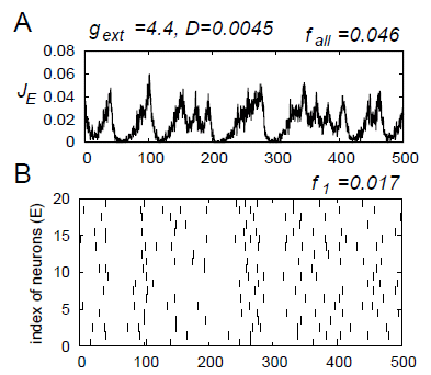
Said in another way - the synchrony is high when the impulse rate is low, see fig. and an animation here. Electrical synapses stabilizes the synchronization. cit. "The oscillations with small amplitudes near the origin corresponded to stochastic synchrony where the ensembleaveraged dynamics shows synchronization in the network but each neuron has a low firing rate and the firing seems to be stochastic. We found stochastic synchrony that corresponded to a chaotic attractor, and we called this phenomenon stochastic synchrony of chaos."

Stochastic synchrony is a phenomenon where the ensemble-averaged dynamics of the network shows synchronization, but the firing rate of each neuron is very low (weak synchronization).
In stochastic synchrony of chaos, the ensemble-averaged dynamics is chaotic. A typical firing pattern of stochastic synchrony of chaos is shown, see Fig. Inhibitory neurons and electrical synapses are important (1).

When stochastic synchrony exists in a network, the contribution of a single neuron in the network to the synchronization is small. Therefore, this neuron might also contribute to form other synchronous networks simultaneously. We get dynamical cell assemblies, reentrant loops in brain a la Edelman, or multichannel neurons. "The question of how a nerve communicates remains unanswered. It is a huge, gaping hole, at the most basic level, in our understanding of how the nervous system works" asks the author.

The dendritic loop/tripartite synapse
There are four types of connections among excitatory and inhibitory assemblies. Are these corresponding to the different brainwave levels? For ex. when visual stimulation was given to cats, oscillations of 40 Hz appeared in the local field potential in the visual cortex and when correlated inputs were given to the receptive fields of each assembly, synchronization among distant (c.7mm) assemblies appeared.
There are also self-oscillating neurons. Autonomous firing.

The modern view holds the neuron as a discrete cell that processes information in more ways than originally envisaged. Intercellular communication by gap junctions, slow electrical potentials, action potentials initiated in dendrites, neuromodulatory effects, extrasynaptic release of neurotransmitters, and information flow between neurons and glia all contribute to information processing. The tripartite synapse is very important.

Dendrites are the analog part of the neurone, and the action potentials are built up in the dendritic tree as an intracellular Ca-wave in form of second messenger.

Collective ion channel gating
Receptor cells make up a syncytium. The response property of the receptor cells, or syncytia, depends on dynamics of collective ion channel gating in the cells, which are 1. periodic, 2. stably chaotic, and 3. unstably chaotic. The dynamics of unstable chaos state synchronizes the potential variation of all receptor cells in the syncytium induced by external stimuli. The syncytium system in the stable and unstable chaos states can detect a weak periodic input signal without assistance of any level of noise (2).

Impulse strength regulation
Then I read that in learning there are rapid shifts of AMPA and NMDA receptors due to exo- and endocytosis, that is communication with the intercellular environment. LTD (and LTP?) is at least partly postsynaptic, and a functional consequence of dendritic protein synthesis is the regulation of glutamate receptor trafficking (3).
Application of amyloid-beta (Alzheimers) promoted endocytosis of NMDA receptors in cortical neurons (4). In addition, neurons from a genetic mouse model of Alzheimer disease expressed reduced amounts of surface NMDA receptors. Reducing amyloid-beta by treating neurons with a gamma-secretase inhibitor restored surface expression of NMDA receptors. Phosphorylation and dephosphorylation seem to be very important, and with it the flux of receptors on/off and the information trafficing. And outside the cell there must be a pool of receptors in off-stage. Also the cytoskeleton is very important, so Ca and Mg (5). The degree of activity of NMDARs is determined in part by extracellular Mg(2+) and by the co-agonists for this receptor, glycine and D-serine (note trans-form). During strong stimulation, a relief of the voltage-dependent block of NMDARs by Mg(2+) provides a positive feedback (5). Alterations in the various balances underlie "meta-plasticity."

So chaos sends the message which the more harmonious states (= slow) detect? In the nerve pulse the solitone is scanning the fibre for disturbances = inputs of information to the system. The harmonious states are the ground states? What makes the information detectable?

Epilepsy shows glutamate synchronize too
Glia and astrocytes make up most of the cells in the brain. It is now becoming clear that these cells make crucial contributions to the formation, operation and adaptation of neural circuitry. The synchrony of abnormally excitable neurons, or even hypersynchrony, is a hallmark in epileptic seizure. Neurons in the brain of individuals with focal epilepsy exhibit sustained discharges, called paroxysmal depolarization shifts. Epileptic discharges are in part initiated by a local depolarization shift that drives groups of neurons into synchronous bursting(11). Unexpected new evidence indicates that glutamate release from glia can generate these events, and may serve to synchronize the activity of neurons (10). Shifts can also be initiated by release of glutamate from extrasynaptic sources or by photolysis of caged Ca2+ in astrocytes. Too big Ca2+-waves are responsible? The mechanisms underlying simultaneous activation of multiple neurons remains still unclear. For epilepsy the astrocytes are very interesting medical targets.

The nonlinear oscillations in two neurons coupled via gap junction in epilepsy, the model of a pair of neurons is derived from Chay model that gives many kinds of abnormal oscillations in excitable neurons by three nonlinear variables dynamics equations. The synchrony between two electrically coupled excitable neurons is found and a theoretical effort is carried out to investigate the chaos in the synchronous oscillations of the membrane potentials by the Lyapunov exponent and the phase portrait. It is shown that synchronous abnormal oscillations of membrane potentials can occur when the coupling strength of gap junction is large enough and the concentration of Ca2 ions does not synchronize while the membrane potentials are synchronous and the coupling mechanism is chaotic (6). It is concluded that the synchrony and the chaos make birth to the new oscillations while disorder process such as epileptic seizure.


A schematic representation of the current and fluxes captured by the Chay 1997 pancreatic beta-cell model. This diagram shows the plasma membrane currents associated with burst and spike oscillations: the fast current, Ifast; the calcium current, I Ca2+ ; the cationic nonselective inward current, INS; the delayed-rectifying K+ current, IK(dr), the calcium-sensitive K+ current, IK(Ca); the ATP-sensitive K+ current, IK(ATP); and the Na+ leak current, INa(L). The ER intracellular Ca2+ store is also shown with its associated transmembrane calcium fluxes: calcium release, Jrel; and calcium uptake by the Ca2+-ATPase, Jpump (7).

Inhibition of impulse
GABA A and GABA B Synapses Play Opposite Roles in Synchronization. They are inhibitory signals(9). Asynchronous release can be seen as a way to produce long-lasting inhibition despite the fast decay time of individual events mediated by GABA A receptors. Asynchronous GABA release produces long-lasting inhibition and accentuates temporal dispersion. Network heterogeneity and synaptic failure play the same role in breaking synchrony.

Myelination
Schwann cells are the glial cells of the peripheral nervous system. They form myelin sheaths, which are essential for saltatory conduction in normal nerves, and promote axonal regeneration in damaged nerves. The transcription factor NF- B, increasingly recognized as a key regulator of developmental and adult neural plasticity, is now reported to be essential for axon myelination as well. What about its function. An isolator, and what more?

Chaos in synchrony of abnormal oscillations
The neurons are connected by both chemical synapses and electrical synapses among the inhibitory neurons. The synchronous firing of neurons in a pulse-coupled neural network composed of excitatory and inhibitory neurons shows that when electrical synapses are introduced, periodically synchronized firing as well as chaotically synchronized firing is widely observed. Moreover, we find stochastic synchrony where the ensemble-averaged dynamics shows synchronization in the network but each neuron has a low firing rate and the firing of the neurons seems to be stochastic. Stochastic synchrony of chaos corresponding to a chaotic attractor is also found (1).

Gamma-frequency (30-70 Hz) oscillations in populations of interneurons may induce synchronous firing in principal neuron networks. Such a role would require that neurons, 1 mm or more apart, are able to synchronize their activity, despite the presence of axonal conduction delays and of the limited axonal spread of many interneurons. Interneuron doublet firing can help to synchronize gamma oscillations, provided that sufficiently many pyramidal neurons are active; gap junctions, between the axons of principal neurons, could contribute to the long-range synchrony of gamma oscillations (in vitro). (Gamma oscillations in isolated networks of tonically excited interneurons, with frequency gated by mutual GABA(A) receptor-mediated IPSPs (interneuron network gamma)). There is simulation and electrophysiological evidence that interneuronal gap junctions (presumably dendritic) can enhance the synchrony of such gamma oscillations (7), in spatially extended interneuron networks. There appears to be a sharp threshold conductance, below which the interneuron dendritic gap junctions do not exert a synchronizing role.

The astroglial syncytium
Glial cells is part of the connective tissue, that made up one single sheat of cells going through the whole body. The sheat has "pockets" for the organs, and fascias make up the "high roads". And there are a lot of fast connections between the cells. Small aqueous pores, the gap junction channels, couple glial astrocytes into an extensive syncytium-like organisation. Astrocytes are the most abundant cell type in the central nervous system (CNS) and increasing evidence now suggests that they play an active role in various brain functions. Astrocytes are involved in the induction and maintenance of the blood brain barrier, as well as the induction and stabilization of neuronal synapses (8).

Astroglial gap junctions are mainly composed of connexin-43 proteins. They provide a pathway for intercellular diffusion of ions and small ((1000 Da) molecules, such as second messengers as Ca2+ and metabolites. Moreover, astrocytes control the extracellular ionic homeostasis, recycle neurotransmitters and painsgiving substance P among others.

Astrocytes become reactive, a process known as reactive gliosis, in CNS pathologies, such as ischemia, neurotrauma or neurodegeneration. Reactive astrocytes seem to be protective at an early stage after neurotrauma but inhibit regeneration later on.

The organisation into multicellular functional units is probably a prerequisite for the participation of astroglial cells in the control of extracellular homeostasis. Waves of increased intracellular Ca2+ concentration can propagate between astrocytes. This is of particular interest, as cytosolic Ca2+ is a second messenger that affects ion channels, carriers, and enzymes and thereby mediates short-, intermediate-, and long-term effects on astroglial function. The modulation of gap junction communication and intra- and intercellular Ca2+ signalling induced by various neuroactive substances may depend on differences in connexin-43 expression, gap junction communication, and Ca2+ signalling in various brain regions.

References
1. Takashi Kanamaru and Kazuyuki Aihara 2008: Stochastic synchrony of chaos in a pulse coupled neural network with both chemical and electrical synapses among inhibitory neurons, Neural Computation, vol.20, no.8 (2008) pp.1951-1972. http://brain.cc.kogakuin.ac.jp/~kanamaru/Chaos/e/sSync/kanamaru-nc2008.pdf

2. Funakubo, H. Kashimori, Y. Kambara, T. 1997: Stable and unstable chaos states of receptor cell syncytium and stochastic resonance without noise. Neural Networks,1997. International Conference on... Publication Date: 9-12 Jun 1997: vol.1: 318-323. http://ieeexplore.ieee.org/xpl/freeabs_all.jsp?tp=&arnumber=611686&isnumber=13361

3. Snyder EM, Philpot BD, Huber KM, Dong X, Fallon JR, Bear MF. 2001: Internalization of ionotropic glutamate receptors in response to mGluR activation.Nat Neurosci. 2001 Nov;4(11):1079-85. http://www.ncbi.nlm.nih.gov/pubmed/11687813">

4. Snyder EM, Nong Y, Almeida CG, Paul S, Moran T, Choi EY, Nairn AC, Salter MW, Lombroso PJ, Gouras GK, Greengard P. 2005: Regulation of NMDA receptor trafficking by amyloid-beta. Nat Neurosci. 2005 Aug;8(8):1051-8. http://www.ncbi.nlm.nih.gov/pubmed/16025111?ordinalpos=38&itool=EntrezSystem2.PEntrez.Pubmed.Pubmed_ResultsPanel.Pubmed_DefaultReportPanel.Pubmed_RVDocSum">endocytosis

5. MacDonald JF, Jackson MF, Beazely MA. 2006: Hippocampal long-term synaptic plasticity and signal amplification of NMDA receptors. Crit Rev Neurobiol. 2006;18(1-2):71-84. http://www.ncbi.nlm.nih.gov/pubmed/17725510?ordinalpos=1&itool=EntrezSystem2.PEntrez.Pubmed.Pubmed_ResultsPanel.Pubmed_DiscoveryPanel.Pubmed_Discovery_RA&linkpos=5&log$=relatedreviews&logdbfrom=pubmed

6. Ge Manling Guo Hongyong Dong Guoya Jia Wenyan Li Ying Sun Minggui Wang Baozhu Yan Weili 2004: The chaos in the synchrony of abnormal oscillations in a pair of neurons coupled via gap junction. Signal Processing, 2004. Proceedings. ICSP '04. 2004 7th International Conference on...vol.3: 2210- 2213 ISBN: 0-7803-8406-7 http://ieeexplore.ieee.org/Xplore/login.jsp?url=http%3A%2F%2Fieeexplore.ieee.org%2Fiel5%2F9834%2F30995%2F01442217.pdf&authDecision=-203

7. Teresa Ree Chay, 1997: Effects of extracellular Calcium on Electrical Bursting and Intracellular and Luminal Calcium Oscillations in Insulin Secreting Pancreatic Beta-Cells, 1997, Biophysical Journal, 73, 1673-1688. PubMed ID: 9284334

8. Nicola J. Allen & Ben A. Barres 2009: Neuroscience: Glia — more than just brain glue. Nature 457, 675-677. http://www.nature.com/nature/journal/v457/n7230/full/457675a.html

9. Martinez D, Montejo N, 2008 A Model of Stimulus-Specific Neural Assemblies in the Insect Antennal Lobe. PLoS Comput Biol 4(8): e1000139. doi:10.1371/journal.pcbi.1000139 http://www.ploscompbiol.org/article/info:doi/10.1371/journal.pcbi.1000139

10. Michael A Rogawski(2005): Astrocytes get in the act in epilepsy. Nature Medicine 11, 919 - 920. http://www.nature.com/nm/journal/v11/n9/abs/nm0905-919.html

11.Guo-Feng Tian et.al. (2005): An astrocytic basis of epilepsy. Nature Medicine 11, 973 - 981. http://www.nature.com/nm/journal/v11/n9/abs/nm1277.html

lördag 18 juli 2009

Modeling at the Gates of the Cell

Got a mail about possibility to win 25000 dollars and there it was. The model of a gap junction. Last year Dr Fleischman received such a prize. He investigated the structure, function, and evolution of membrane proteins associated with hereditary hearing loss and neurodegenerative diseases, cancer, and bacterial drug resistance. He ties to understand the mechanistic relationship between molecular structure and function in human health and disease.

He says: Lack of an atomic resolution structure of the gap junction has made it extremely difficult to conduct biochemical investigations within a consistent framework ... it becomes clear that we have an unsatisfactory picture of the possible structural motifs found in membrane proteins. This limited viewpoint raises the question: Can we apply computational modeling to provide additional insight into the relationship between structure and function in membrane proteins? Experimental data along with evolutionary analysis may help us bridge the gap in our structural understanding of membrane proteins and provide structural models.

An updated overview of current knowledge of connexins and their interacting proteins and connexin modulation, disease and tumorigenesis is made by Dbouk et.al. 2009.

The cores of membrane proteins are much more evolutionarily conserved than their peripheries. The reason for this is simple: Mutation at the core of the protein is much more likely to disrupt the protein structure than one in a lipid-facing position, and would be eliminated by the forces of natural selection. Evolutionary conservation could therefore distinguish the parts of each helix that face the lipid from those that face the core of the protein.

Gap junctions links the cytoplasms of neighboring cells in mammalian tissues and allows the cells to transfer metabolites and signals. It is a critical component in cellular signaling of many tissues, and numerous mutations in its membrane domain have been implicated in hereditary hearing loss, neurodegenerative disease, and other genetic diseases.


Molecular organization of a recombinant gap junction channel. The approximate boundaries for the membrane bilayers (M), extracellular gap (E), and the cytoplasmic space (C) are indicated.


(a) A top view looking toward the extracellular gap and (b) a side view of part of the 3D map of a recombinant gap junction channel. The 24 well-resolved rod-shaped features reveal the packing of the transmembrane alpha helices, and four have been arbitrarily labeled A, B, C, and D.

Gap junction channels are formed by the end-to-end docking of two hemichannels or connexons = hexamers, from adjacent membranes, each of which displayed 24 rods of density in the membrane interior. Each connexon comprises six connexin subunits, proteins which are encoded by ~20 isoforms in the human genome. All connexins contain four transmembrane (TM)4 segments (M1-M4), whose N and C termini are located in the cytoplasm. The extracellular aspect of the hemichannel is composed of two extracellular loops (E1 and E2) from each connexin monomer. A long-awaited high-resolution structure of a connexin channel was published recently (Maeda et al. 2009).

Gating
The gating mechanism that achieves opening is voltage and pH sensitive, and protein phosphorylation - sensitive, and requires extracellular calcium ion in the millimolar range to remain closed at normal resting potentials. Unapposed connexin hemichannels exhibit robust closure in response to membrane hyperpolarization and extracellular calcium. This form of gating, termed "loop gating," is largely responsible for regulating hemichannel opening. The molecular components and structural rearrangements underlying loop gating remain unknown. Metal bridges can lock up the gate. Sulfhydryl groups, the contribution from disulfide bond formation and other residues that coordinate metal ions with high affinity are other candidates. Metal ions access the cysteine side chains through the open pore and that closure of the loop gate involves movement of the TM1/E1 region that results in local narrowing of the large aqueous connexin pore. The loop gate must also be able to open when docked to another hemichannel in the junctional configuration. The relationship between loop gating and the mechanism/structure of hemichannel docking is unclear. Perhaps the mechanism of loop gate formation is different in connexin isoforms? An alternative possibility is that the binding and the opening are two distinct processes that are not obligatorily linked. This scenario permits docking without opening of the pore; docking would enable opening but not require it. In this case, the structural elements involved in the two processes would need to be distinct.

What about the basis of the voltage sensitivity of the loop gate? Connexin channels have at least one other gating mechanism, known as Vj gating, which closes the channels to a substate and is well characterized at the single-channel level. The sensor for this type of gating was within the pore, composed of the amino-terminal domain of the protein, which when the gate is open is folded into the lumen of pore against TM1, forming the pore wall in the cytoplasmic end of the pore. The suggestion is that in response to an appropriate electric field, these domains peel off the pore wall and move toward the cytoplasm to collapse into an aggregate that largely occludes the lumen. This is a unique voltage-dependent gating mechanism, operating at the opposite end of the pore from the loop gate. Data suggest that the voltage sensors of the two mechanisms are in series in the lumen of the pore, and that the sensitivity of each to applied voltage changes with the position of the other gate.

The cytoplasmic N-termini of connexins have been implicated in protein trafficking, oligomerization and channel gating. Mutants containing nine or more N-terminal amino acids form gap junction plaques. This N-terminal peptide is predominantly {alpha}-helical. The {alpha}-helical structure of the connexin37 N terminus may be dispensable for protein localization, but it is required for channel and hemichannel function. As much as half the length of the connexin N-terminus can be deleted without affecting formation of gap junction plaques, but an intact N-terminus is required for hemichannel gating and intercellular communication. The NT does not need to be the gate itself. Loss of conductance in the NT deletion mutants might also result if an intact NT is required for the other gates to be operative.


Superposition of cross sections (red and blue) within the hydrophobic region of the two hexameric connexons forming the gap junction channel. The shaded regions identify three possible boundaries for the connexin subunit. With reference to the labels in Fig. 3a, the models show (a) a bundle of four alpha helices (A'DCB'), (b) a check-mark arrangement (ABCD), and (c) a zigzag pattern (BCDA'). White arrows identify axes of symmetry that are in plane, noncrystallographic, and twofold.


The model structure of the gap junction membrane domain helps to explain the differential effect of disease-causing and benign polymorphisms.

Fleishmans group made a model of a gap junction domain. The model structure helped identify positions on adjacent helices where second-site mutations restored membrane localization, revealing possible interactions between residue pairs. We thus identified two putative salt bridges and one pair involved in packing interactions in which one disease-causing mutation suppressed the effects of another. These results seem to reveal interactions that apparently stabilize contacts between TM helices in connexins and suggest that abrogation of such interactions bring about some of the effects of disease-causing mutations.

The stabilizing structure is a triad of charged positions (salt-bridges), the first two occupied by basic residues and the last by an acidic residue, are highly conserved throughout all connexins. In theory, the Arg and Glu residues, which are reciprocally charged and are near the water-filled pore lumen, could be involved in stabilizing electrostatic interactions; it has been estimated that salt bridges embedded in water can add nearly 1 kcal/mol to protein stability. Additional forces contribute to stabilization (no salt bridge).

Although wild-type connexins are membrane-localized, our images show fluorescence also outside the membrane, even in wild-type connexin. Localization of wild-type connexins outside the membrane, in addition to the existence of gap junction plaques, have been observed in other studies involving overexpressed connexins, and it has been suggested that the cytoplasmic fraction of the protein is at least in part localized in aggresomes. The important point to notice is that, along with the localization of some of the protein in the cytoplasm, wild-type and doubly mutated connexins are localized in the plasma membrane, whereas the single mutants are not.

Tight junctions
Unger et.al. write:... the protein density that formed the extracellular vestibule provided a tight seal to exclude the exchange of substances with the extracellular milieu.

Remembers me of tight junctions. In brain we have the brainbarrier, in nerves we have a tight junction seal, in liver and heart too. What kind of purpose do that have? Sorting big molecules out only?

From the rewiev: The adherens and tight junctions regulates paracellular permeability (barrier function) and cell polarity, among other functions. Gap junctions may even regulate the expression and function of tight junction proteins. The tight junction membrane-associated guanylate kinase protein is involved in the organization and trafficking of gap junctions, and mediates the delivery of Cx43 from a lipid raft domain to gap junctional plaques. Cx32 can participate in the formation of functional tight junctions and in actin organization. Interactions of connexins with the actin cytoskeleton and associated proteins serve to stabilize gap junctions at the plasma membrane.

Among newly-discovered interacting connexin partners, plasma membrane ion channels, membrane transport proteins and receptors have been shown to interact directly or indirectly with connexins, and these include aquaporin-0 and acetylcholine receptors, among others. The calcium/calmodulin-dependant kinase II (CaMKII) interacts with and phosphorylates. Other interactions include cholesterol, COX-2 and heat shock protein 90 (Hsp90) and translocase of the outer mitochondrial membrane.

Interaction of connexins with CaMKII may have a general regulatory role in neuronal signal transmission, with a role in electrical coupling in addition to the defined role of CaMKII in chemical synaptic transmission.

Recent studies have revealed complex translational and post-translational mechanisms that regulate connexin synthesis, maturation, membrane transport and degradation that in turn modulate gap junction intercellular communication. With the growing myriad of connexin interacting proteins, including cytoskeletal elements, junctional proteins, and enzymes, gap junctions are now perceived, not only as channels between neighboring cells, but as signaling complexes that regulate cell function and transformation. They exert their effects on proliferation and other aspects of life and death of the cell through mostly-undefined mechanisms.

An array of studies illustrated the important contribution of GJIC to developmental and regulatory events such as embryonic growth, bone modeling, alveolar differentiation, central nervous system signaling and neural function in the developing central nervous system, also in maintenance of tissue homeostasis through processes such as synchronization.

Tis is very interesting for my meridians. Very, very much.

References
Sarel J. Fleishman, Adi D. Sabag, Eran Ophir, Karen B. Avraham, and Nir Ben-Tal 2006: The Structural Context of Disease-causing Mutations in Gap Junctions. J. Biol. Chem., Vol. 281, Issue 39, 28958-28963, September 29, 2006. http://www.jbc.org/cgi/content/full/281/39/28958

Sarel Fleishman 2008: Modeling at the Gates of the Cell.
http://www.sciencemag.org/feature/data/prizes/ge/2008/fleishman.dtl

Vinzenz M. Unger, Nalin M. Kumar, Norton B. Gilula, Mark Yeager 1999: Three-Dimensional Structure of a Recombinant Gap Junction Membrane Channel. Science 19 February 1999: Vol. 283. no. 5405, pp. 1176 - 1180. http://www.sciencemag.org/cgi/content/full/283/5405/1176

A. L. Harris, 2009: Gating on the outside. J. Gen. Physiol., June 1, 2009; 133(6): 549 - 553. http://jgp.rupress.org/cgi/content/full/133/6/549

Maeda, S., S. Nakagawa, M. Suga, E. Yamashita, A. Oshima, Y. Fujiyoshi, and T. Tsukihara. 2009. Structure of the connexin 26 gap junction channel at 3.5 A resolution. Nature. 458:597–602.

Oshima, A., Doi, T., Mitsuoka, K., Maeda, S. and Fujiyoshi, Y. (2003). Roles of Met-34, Cys-64, and Arg-75 in the assembly of human connexin 26: Implication for key amino acid residues for channel formation and function. J. Biol. Chem. 278, 1807-1816. http://www.jbc.org/cgi/content/abstract/278/3/1807?ijkey=7588b0090e287999b2f3cc079c2c694006d66731&keytype2=tf_ipsecsha

J. W. Kyle, P. J. Minogue, B. C. Thomas, D. A. L. Domowicz, V. M. Berthoud, D. A. Hanck, and E. C. Beyer (2008): An intact connexin N-terminus is required for function but not gap junction formation. J. Cell Sci. 121, 2744-2750 http://jcs.biologists.org/cgi/content/full/121/16/2744

V. K. Verselis, M. P. Trelles, C. Rubinos, T. A. Bargiello, and M. Srinivas (2009): Loop Gating of Connexin Hemichannels Involves Movement of Pore-lining Residues in the First Extracellular Loop Domain. J. Biol. Chem. 284, 4484-4493.

torsdag 16 juli 2009

Ring hexamers

Matti Pitkänen wrote in his blog some interesting comment: Ring hexamers bring in mind the crucial role of aromatic cycles in TGD inspired model of DNA as topological quantum computer which leads also to a model of ADP→ ATP transition involving reconnection of magnetic flux tubes and having also information theoretic interpretation as a change of the topology of the braid structure defining topological quantum computer program. Magnetic flux tubes carrying dark electrons begin from these and can end up to other biomolecules or water. Just a guess: could they end on ring hexamers?

Earlier I had read about ring hexamers giving rise to a magnetic field. Ring hexamers are carbon rings. And they can give rise to coronenes with a self-organizing capacity, supramolecules, chrystals and nanotubules in artificial conditions. Also fullerenes and nanotubules give rise to fluorescent fields, perhaps also magnetic, as I heard from a scientist in Umeå, Sweden.

Nighttime view of Saturn's north pole shows a bizarre six-sided hexagon feature encircling the entire north pole, a long-lived feature. A second hexagon, significantly darker is also visible. The red color indicates the amount of 5-micron wavelength radiation, or heat, generated in the warm interior of Saturn that escapes the planet. The hexagon is similar to Earth's polar vortex, which has winds blowing in a circular pattern around the polar region. On Saturn, the vortex has a hexagonal rather than circular shape. The hexagon is nearly 25,000 kilometers (15,000 miles) across. Nearly four Earths could fit inside it. This is a very strange feature, lying in a precise geometric fashion with six nearly equally straight sides, and anything like this has never been seen on any other planet.A system of clouds lies within the hexagon. The clouds appear to be whipping around the hexagon like cars on a racetrack. The hexagon images and movie. Image credit: NASA/JPL/University of Arizona


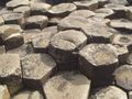
Hexamers are very common in nature. The bees use it in their hives. Giant's Causeway has volcanic basalt columns forming stepping stones that lead from the cliff foot and disappear under the sea. Most of the columns are hexagonal in form.


Then I remembered that some receptors have six proteine parts or helices, as acuaporines and gap junctions. Ordered in a ring structure like a hexamer. And the receptors are ordered in clusters, topologically arranged often. The neurons can even distinguish between left side of body and right side. The neurons/receptors are asymmetric.

About channels and their ligands(Ashcroft 2000)
Ligand-gated channels are generally named after the ligand that gates them. These may be extracellular, as in the case of the neurotransmitters acetylcholine and glycine, or intracellular such as cyclic AMP, Ca2+, and ATP. Binding of the ligand to one or more specific sites on the channel protein produce a conformational change that allosterically opens the ion pore, but sometimes close it.

At the resting potential of the cell, most voltage-gated channels are closed. When the membrane potential is changed, however. the channel undergoes a series of conformational changes that result in the opening of the channel pore. Also hyperpolarized gating exists.

The gating of almost all ion channels is subject to modulation by one or
more of a wide range of substances. These include monovalent and divalent cations (such as H- and Ca2-), metabolites such as ATP and MgADP, fatty acids, phosphorylation, GTP-binding proteins, and even gases (such as oxygen). The voltage dependence of activation is shifted to more negative membrane potentials in the presence of intracellular Ca2+. Sometimes the distinction between a channel modulator and the principal ligand seems merely semantic. Many hormones and neurotransmitters mediate their effects on ion channels indirectly by the activation of some second messenger system that modulates ion channel activity.

Chemotactic receptors
One of the best understood regions of cytoplasm is that associated with the cluster of chemotactic receptors in the plasma membrane of E. coli. Bacterial chemotaxis involves a phospho-relay system. This signaling complex transmits information from outside into the cell in the form of a phosphorylation signal that regulates flagellar rotation. The receptors are believed to exist in thermal equilibrium between two or more conformational states, and the output of the complex, usually measured in terms of phosphorylation levels is related to this equilibrium. The complex produces an amplified output proportional to the rate of change of attractant concentration.

The eukaryotic cell exhibits compartmentalization of functions to various membrane-bound organelles and to specific domains within each membrane.

An atomic level structure for a lattice of serine (Tsr) receptors are proposed. A unique feature of this model is that it creates a small compartment between the plasma membrane and an extended hexagonal lattice of the signaling proteins CheA and CheW. The compartment is not closed, and should be freely accessible to cytoplasmic proteins diffusing in from the lateral borders or through 10 nm diameter pores in the hexagonal lattice. This minute volume of bacterial cytoplasm will be highly enriched in two diffusible proteins = two enzymes that control the methylation of chemotactic receptors at sites in the middle of the receptor tails and have also been shown to carry binding sites for the C-termini of the receptor tails. It seems reasonable to suppose that they will accumulate in the small compartment because of this binding, and that their elevated concentrations in that privileged volume will facilitate the methylation and demethylation reactions.


The dynamics of the situation lead to the spontaneous emergence of order in the receptor lattice, such that receptors with 4 methyl groups (fully methylated) and receptors with 0 methyl groups (fully unmethylated) tend to lie next to each other in the array. The spontaneous emergence of order within a stochastically fluctuating field of allosteric proteins is an intriguing and potentially important phenomenon. By assigning different values of coupling energy to these different protein interactions and then examining the consequences for an array with the geometry of the receptor lattice the simulation model will be better.

This is fantastic. Sounds very TGD:ic in my ears. And reflexologic with all those geometric topologies.

Activity spread
The remarkable sensitivity and range of response of bacterial chemotaxis might depend on the clustering of chemotactic receptors on the surface of the bacterium. When a ligand bound to a receptor, the change in activity might propagate to neighbouring receptors in a cluster. Activity was spread by energetic exchanges between proteins in a two-dimensional lattice could be likened to the interactions of magnetic dipoles in a spin glass, resulting in digital receptors, also affected of the state of neighbours, "coupling energy".

Snapshot of an array of receptors showing a spread of activity. The receptors are portrayed as hexagons in an instantaneous snapshot showing their conformation as being either inactive (white) or active (orange).Bray et.al.1998.


Freeze fracture replica of two gap junctional plaques made up of a cluster of connexons (Ashcroft 2000),

Projected density map showing six-folded symmetry, rat Cx43 (Asgcroft 2000). See the neighbouring cells that are very sensitive to electric and magnetic changes. These cells are modulators (eg. their peptides).

Fluid mosaic membrane
It is assumed usually that the lipid membrane acts as an anchor for the proteins and has no independent function. However, membranes contain hundreds of different lipids (depicted in different colors in Fig.) which are different with respect to their physical properties (for example charge and size). One may wonder why nature has put so much effort into creating all these lipids if there is no functional purpose linked to their diversity. It is (for instance) known that bacteria change their lipid composition in response to changes in environmental conditions (e.g. different temperature of growth). Why is that, asks Heimburg?

Experiments and theoretical studies on model membranes, however, lead to the conclusion that the "fluid mosaic"-model requires major revision.


Modified version of the famous picture of the plasma membrane (Singer and Nicolson, 1972). Lipids (shown in different colors) are not uniformly distributed all over the membrane, but form domains of varying composition. Integral and peripheral proteins are depicted in green.

An important observation in a Monte-Carlo simulation (increasing temp.) is the formation of domains made of groups of gel or fluid lipids. Gel is in patches.
If proteins bind to a membrane with lipid domains, these proteins will be unevenly distributed on the membrane and the local protein concentration on the membrane surface may be quite variable. Membranes become soft and flexible in the transition regime.

Wave genome and phantom DNA?
Matti Pitkänen write: "Gariaev and collaborators have introduced the notion of wave genome requiring the coding of DNA sequences to temporal patterns of coherent em fields forming a bio-hologram representing geometric information about the organism. Code could mean that nucleotide is represented by a characteristic rotation angle for the polarization plane of linearly polarized laser radiation scattering from it. This kind rotation is known to be induced by chromosomes. Gariaev had photographs produced by the scattering of ordinary light on DNA, showing amazing "memory". In TGD framework these photographs could be interpreted as photographs of wormhole magnetic flux tubes containing dark matter."

The ability of this kind of light to induce gene expression in another organisms provided the modulated polarization pattern corresponds to an "address" characterizing the organism, and the formation of images of what is believed to be DNA sample itself and of the objects of environment by DNA sample in a cell irradiated by ordinary light in UV-IR range. This means that it works all ways, from within the cell reflekted out, or from outside reflected inside? A truly reflexologic picture.

What is even nastier is that in this way we can show how the DNA influences the cell membranes. And suddenly all those magnetic flux tubes and braidings makes sense. Even the magnetic body doesn't sound so peculiar. Biology is really quantum biology? As Matti said once "The guy was rightafter all?"

I can see how the chromosomes induces magnetic flux tubes, that are fastened to the receptors tails. And also to receptors on other membranes. By that neighbour-effect is then patches built on the membrane. That is phase-transitions, regulating the activity of the domain by inducing a gel. The signal reaching the membrane can be magnetic, electric or chemical (ionic) with hexamers as a vey important part. In fact life is about carbon, and carbon is about organic chemistry. In quantum physics there are the holy trinity, and a hierarchy of that forms hexamers.

Perhaps Matti he himself will explain these things in more detail to us when he has time for it. We are waiting for that impatiently.

What has all this in common with the Saturn pole? It is ice, or water. More about that later.

References:

Acshcroft 2000: Ion channels and disease.

Bray et.al. 1998, www.pdn.cam.ac.uk/groups/comp-cell/Papers/Bray98a.pdf

Crystal structure of a molecular hexagon composed of hexagonal aromatic rings reported by Müllen and coworkers in Chem. Eur. J., 2000, 1834-1839.
http://en.wikipedia.org/wiki/Coronene

Cassini Images Bizarre Hexagon on Saturn 03.27.07, http://www.nasa.gov/mission_pages/cassini/media/cassini-20070327.html

Heimburg,T. 1998?: AG 012 - Membrane Thermodynamics. The fluid-mosaic model. Fluctuations and Domains: Thermodynamic properties and heterogeneity of membrane assemblies. http://membranes.nbi.dk/presentation-english/News_engl.html

Maddock, J. R., & Shapiro, L. (1993). Polar location of the chemoreceptor complex in the Escherichia coli cell. Science 259, 1717-1723. http://www.sciencemag.org/cgi/content/abstract/259/5102/1717

Pitkänen, M. 2008: Has dark matter at the magnetic flux tubes been photographed? http://matpitka.blogspot.com/2008/05/has-dark-matter-at-magnetic-flux-tubes.html.

Receptor Clusters, 2006. Bray Group: Computer Models of Bacterial chemotaxis, Physiology, Development and Neuroscience, University of Cambridge, http://www.pdn.cam.ac.uk/groups/comp-cell/Cluster.html