tisdag 21 juli 2009

Chaos and synchrony

Cells are not solitary individuals. They function in an orchestra, a syncytium. They are social and talk to each other. They can sing, and they can scream or make noise, be rebellious. And I have always thought that the song is the good one, that which organize and regulate the cells. That the noise is the bad guy, that brings pain and sickness. Then I read that the noise is the regulating one. In biology, nothing is what it used to be any more. The world has turned upside down. And we must try to extract the truth from that resulting mess.


That the noise is the regulating force, or the basic information, is true for the nerve pulse in the solitone. Also in the brainstem the RAS-network inhibite strongly the impulses from the body, and a further inhibition is done by the blood-brain barrier (tight junction) and modulation by neurotransmitters and peptides. Modulation change the brainwaves and gives them a higher amplitude. Most of the modulation is done in the midbrain in thalamus-hypothalamus region as emotions. Cortical modulation is done by neurotransmitters. The "driving force" is glutamat, and inhibitory force is GABA.

Said in another way - the synchrony is high when the impulse rate is low, see fig. and an animation here. Electrical synapses stabilizes the synchronization. cit. "The oscillations with small amplitudes near the origin corresponded to stochastic synchrony where the ensembleaveraged dynamics shows synchronization in the network but each neuron has a low firing rate and the firing seems to be stochastic. We found stochastic synchrony that corresponded to a chaotic attractor, and we called this phenomenon stochastic synchrony of chaos."

Stochastic synchrony is a phenomenon where the ensemble-averaged dynamics of the network shows synchronization, but the firing rate of each neuron is very low (weak synchronization).
In stochastic synchrony of chaos, the ensemble-averaged dynamics is chaotic. A typical firing pattern of stochastic synchrony of chaos is shown, see Fig. Inhibitory neurons and electrical synapses are important (1).
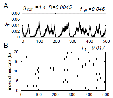
When stochastic synchrony exists in a network, the contribution of a single neuron in the network to the synchronization is small. Therefore, this neuron might also contribute to form other synchronous networks simultaneously. We get dynamical cell assemblies, reentrant loops in brain a la Edelman, or multichannel neurons. "The question of how a nerve communicates remains unanswered. It is a huge, gaping hole, at the most basic level, in our understanding of how the nervous system works" asks the author.

The dendritic loop/tripartite synapse
There are four types of connections among excitatory and inhibitory assemblies. Are these corresponding to the different brainwave levels? For ex. when visual stimulation was given to cats, oscillations of 40 Hz appeared in the local field potential in the visual cortex and when correlated inputs were given to the receptive fields of each assembly, synchronization among distant (c.7mm) assemblies appeared.
There are also self-oscillating neurons. Autonomous firing.

The modern view holds the neuron as a discrete cell that processes information in more ways than originally envisaged. Intercellular communication by gap junctions, slow electrical potentials, action potentials initiated in dendrites, neuromodulatory effects, extrasynaptic release of neurotransmitters, and information flow between neurons and glia all contribute to information processing. The tripartite synapse is very important.

Dendrites are the analog part of the neurone, and the action potentials are built up in the dendritic tree as an intracellular Ca-wave in form of second messenger.

Collective ion channel gating
Receptor cells make up a syncytium. The response property of the receptor cells, or syncytia, depends on dynamics of collective ion channel gating in the cells, which are 1. periodic, 2. stably chaotic, and 3. unstably chaotic. The dynamics of unstable chaos state synchronizes the potential variation of all receptor cells in the syncytium induced by external stimuli. The syncytium system in the stable and unstable chaos states can detect a weak periodic input signal without assistance of any level of noise (2).

Impulse strength regulation
Then I read that in learning there are rapid shifts of AMPA and NMDA receptors due to exo- and endocytosis, that is communication with the intercellular environment. LTD (and LTP?) is at least partly postsynaptic, and a functional consequence of dendritic protein synthesis is the regulation of glutamate receptor trafficking (3).
Application of amyloid-beta (Alzheimers) promoted endocytosis of NMDA receptors in cortical neurons (4). In addition, neurons from a genetic mouse model of Alzheimer disease expressed reduced amounts of surface NMDA receptors. Reducing amyloid-beta by treating neurons with a gamma-secretase inhibitor restored surface expression of NMDA receptors. Phosphorylation and dephosphorylation seem to be very important, and with it the flux of receptors on/off and the information trafficing. And outside the cell there must be a pool of receptors in off-stage. Also the cytoskeleton is very important, so Ca and Mg (5). The degree of activity of NMDARs is determined in part by extracellular Mg(2+) and by the co-agonists for this receptor, glycine and D-serine (note trans-form). During strong stimulation, a relief of the voltage-dependent block of NMDARs by Mg(2+) provides a positive feedback (5). Alterations in the various balances underlie "meta-plasticity."

So chaos sends the message which the more harmonious states (= slow) detect? In the nerve pulse the solitone is scanning the fibre for disturbances = inputs of information to the system. The harmonious states are the ground states? What makes the information detectable?

Epilepsy shows glutamate synchronize too
Glia and astrocytes make up most of the cells in the brain. It is now becoming clear that these cells make crucial contributions to the formation, operation and adaptation of neural circuitry. The synchrony of abnormally excitable neurons, or even hypersynchrony, is a hallmark in epileptic seizure. Neurons in the brain of individuals with focal epilepsy exhibit sustained discharges, called paroxysmal depolarization shifts. Epileptic discharges are in part initiated by a local depolarization shift that drives groups of neurons into synchronous bursting(11). Unexpected new evidence indicates that glutamate release from glia can generate these events, and may serve to synchronize the activity of neurons (10). Shifts can also be initiated by release of glutamate from extrasynaptic sources or by photolysis of caged Ca2+ in astrocytes. Too big Ca2+-waves are responsible? The mechanisms underlying simultaneous activation of multiple neurons remains still unclear. For epilepsy the astrocytes are very interesting medical targets.

The nonlinear oscillations in two neurons coupled via gap junction in epilepsy, the model of a pair of neurons is derived from Chay model that gives many kinds of abnormal oscillations in excitable neurons by three nonlinear variables dynamics equations. The synchrony between two electrically coupled excitable neurons is found and a theoretical effort is carried out to investigate the chaos in the synchronous oscillations of the membrane potentials by the Lyapunov exponent and the phase portrait. It is shown that synchronous abnormal oscillations of membrane potentials can occur when the coupling strength of gap junction is large enough and the concentration of Ca2 ions does not synchronize while the membrane potentials are synchronous and the coupling mechanism is chaotic (6). It is concluded that the synchrony and the chaos make birth to the new oscillations while disorder process such as epileptic seizure.


A schematic representation of the current and fluxes captured by the Chay 1997 pancreatic beta-cell model. This diagram shows the plasma membrane currents associated with burst and spike oscillations: the fast current, Ifast; the calcium current, I Ca2+ ; the cationic nonselective inward current, INS; the delayed-rectifying K+ current, IK(dr), the calcium-sensitive K+ current, IK(Ca); the ATP-sensitive K+ current, IK(ATP); and the Na+ leak current, INa(L). The ER intracellular Ca2+ store is also shown with its associated transmembrane calcium fluxes: calcium release, Jrel; and calcium uptake by the Ca2+-ATPase, Jpump (7).

Inhibition of impulse
GABA A and GABA B Synapses Play Opposite Roles in Synchronization. They are inhibitory signals(9). Asynchronous release can be seen as a way to produce long-lasting inhibition despite the fast decay time of individual events mediated by GABA A receptors. Asynchronous GABA release produces long-lasting inhibition and accentuates temporal dispersion. Network heterogeneity and synaptic failure play the same role in breaking synchrony.

Myelination
Schwann cells are the glial cells of the peripheral nervous system. They form myelin sheaths, which are essential for saltatory conduction in normal nerves, and promote axonal regeneration in damaged nerves. The transcription factor NF- B, increasingly recognized as a key regulator of developmental and adult neural plasticity, is now reported to be essential for axon myelination as well. What about its function. An isolator, and what more?

Chaos in synchrony of abnormal oscillations
The neurons are connected by both chemical synapses and electrical synapses among the inhibitory neurons. The synchronous firing of neurons in a pulse-coupled neural network composed of excitatory and inhibitory neurons shows that when electrical synapses are introduced, periodically synchronized firing as well as chaotically synchronized firing is widely observed. Moreover, we find stochastic synchrony where the ensemble-averaged dynamics shows synchronization in the network but each neuron has a low firing rate and the firing of the neurons seems to be stochastic. Stochastic synchrony of chaos corresponding to a chaotic attractor is also found (1).

Gamma-frequency (30-70 Hz) oscillations in populations of interneurons may induce synchronous firing in principal neuron networks. Such a role would require that neurons, 1 mm or more apart, are able to synchronize their activity, despite the presence of axonal conduction delays and of the limited axonal spread of many interneurons. Interneuron doublet firing can help to synchronize gamma oscillations, provided that sufficiently many pyramidal neurons are active; gap junctions, between the axons of principal neurons, could contribute to the long-range synchrony of gamma oscillations (in vitro). (Gamma oscillations in isolated networks of tonically excited interneurons, with frequency gated by mutual GABA(A) receptor-mediated IPSPs (interneuron network gamma)). There is simulation and electrophysiological evidence that interneuronal gap junctions (presumably dendritic) can enhance the synchrony of such gamma oscillations (7), in spatially extended interneuron networks. There appears to be a sharp threshold conductance, below which the interneuron dendritic gap junctions do not exert a synchronizing role.

The astroglial syncytium
Glial cells is part of the connective tissue, that made up one single sheat of cells going through the whole body. The sheat has "pockets" for the organs, and fascias make up the "high roads". And there are a lot of fast connections between the cells. Small aqueous pores, the gap junction channels, couple glial astrocytes into an extensive syncytium-like organisation. Astrocytes are the most abundant cell type in the central nervous system (CNS) and increasing evidence now suggests that they play an active role in various brain functions. Astrocytes are involved in the induction and maintenance of the blood brain barrier, as well as the induction and stabilization of neuronal synapses (8).

Astroglial gap junctions are mainly composed of connexin-43 proteins. They provide a pathway for intercellular diffusion of ions and small ((1000 Da) molecules, such as second messengers as Ca2+ and metabolites. Moreover, astrocytes control the extracellular ionic homeostasis, recycle neurotransmitters and painsgiving substance P among others.

Astrocytes become reactive, a process known as reactive gliosis, in CNS pathologies, such as ischemia, neurotrauma or neurodegeneration. Reactive astrocytes seem to be protective at an early stage after neurotrauma but inhibit regeneration later on.

The organisation into multicellular functional units is probably a prerequisite for the participation of astroglial cells in the control of extracellular homeostasis. Waves of increased intracellular Ca2+ concentration can propagate between astrocytes. This is of particular interest, as cytosolic Ca2+ is a second messenger that affects ion channels, carriers, and enzymes and thereby mediates short-, intermediate-, and long-term effects on astroglial function. The modulation of gap junction communication and intra- and intercellular Ca2+ signalling induced by various neuroactive substances may depend on differences in connexin-43 expression, gap junction communication, and Ca2+ signalling in various brain regions.

References
1. Takashi Kanamaru and Kazuyuki Aihara 2008: Stochastic synchrony of chaos in a pulse coupled neural network with both chemical and electrical synapses among inhibitory neurons, Neural Computation, vol.20, no.8 (2008) pp.1951-1972. http://brain.cc.kogakuin.ac.jp/~kanamaru/Chaos/e/sSync/kanamaru-nc2008.pdf

2. Funakubo, H. Kashimori, Y. Kambara, T. 1997: Stable and unstable chaos states of receptor cell syncytium and stochastic resonance without noise. Neural Networks,1997. International Conference on... Publication Date: 9-12 Jun 1997: vol.1: 318-323. http://ieeexplore.ieee.org/xpl/freeabs_all.jsp?tp=&arnumber=611686&isnumber=13361

3. Snyder EM, Philpot BD, Huber KM, Dong X, Fallon JR, Bear MF. 2001: Internalization of ionotropic glutamate receptors in response to mGluR activation.Nat Neurosci. 2001 Nov;4(11):1079-85. http://www.ncbi.nlm.nih.gov/pubmed/11687813">

4. Snyder EM, Nong Y, Almeida CG, Paul S, Moran T, Choi EY, Nairn AC, Salter MW, Lombroso PJ, Gouras GK, Greengard P. 2005: Regulation of NMDA receptor trafficking by amyloid-beta. Nat Neurosci. 2005 Aug;8(8):1051-8. http://www.ncbi.nlm.nih.gov/pubmed/16025111?ordinalpos=38&itool=EntrezSystem2.PEntrez.Pubmed.Pubmed_ResultsPanel.Pubmed_DefaultReportPanel.Pubmed_RVDocSum">endocytosis

5. MacDonald JF, Jackson MF, Beazely MA. 2006: Hippocampal long-term synaptic plasticity and signal amplification of NMDA receptors. Crit Rev Neurobiol. 2006;18(1-2):71-84. http://www.ncbi.nlm.nih.gov/pubmed/17725510?ordinalpos=1&itool=EntrezSystem2.PEntrez.Pubmed.Pubmed_ResultsPanel.Pubmed_DiscoveryPanel.Pubmed_Discovery_RA&linkpos=5&log$=relatedreviews&logdbfrom=pubmed

6. Ge Manling Guo Hongyong Dong Guoya Jia Wenyan Li Ying Sun Minggui Wang Baozhu Yan Weili 2004: The chaos in the synchrony of abnormal oscillations in a pair of neurons coupled via gap junction. Signal Processing, 2004. Proceedings. ICSP '04. 2004 7th International Conference on...vol.3: 2210- 2213 ISBN: 0-7803-8406-7 http://ieeexplore.ieee.org/Xplore/login.jsp?url=http%3A%2F%2Fieeexplore.ieee.org%2Fiel5%2F9834%2F30995%2F01442217.pdf&authDecision=-203

7. Teresa Ree Chay, 1997: Effects of extracellular Calcium on Electrical Bursting and Intracellular and Luminal Calcium Oscillations in Insulin Secreting Pancreatic Beta-Cells, 1997, Biophysical Journal, 73, 1673-1688. PubMed ID: 9284334

8. Nicola J. Allen & Ben A. Barres 2009: Neuroscience: Glia — more than just brain glue. Nature 457, 675-677. http://www.nature.com/nature/journal/v457/n7230/full/457675a.html

9. Martinez D, Montejo N, 2008 A Model of Stimulus-Specific Neural Assemblies in the Insect Antennal Lobe. PLoS Comput Biol 4(8): e1000139. doi:10.1371/journal.pcbi.1000139 http://www.ploscompbiol.org/article/info:doi/10.1371/journal.pcbi.1000139

10. Michael A Rogawski(2005): Astrocytes get in the act in epilepsy. Nature Medicine 11, 919 - 920. http://www.nature.com/nm/journal/v11/n9/abs/nm0905-919.html

11.Guo-Feng Tian et.al. (2005): An astrocytic basis of epilepsy. Nature Medicine 11, 973 - 981. http://www.nature.com/nm/journal/v11/n9/abs/nm1277.html

lördag 18 juli 2009

Modeling at the Gates of the Cell

Got a mail about possibility to win 25000 dollars and there it was. The model of a gap junction. Last year Dr Fleischman received such a prize. He investigated the structure, function, and evolution of membrane proteins associated with hereditary hearing loss and neurodegenerative diseases, cancer, and bacterial drug resistance. He ties to understand the mechanistic relationship between molecular structure and function in human health and disease.

He says: Lack of an atomic resolution structure of the gap junction has made it extremely difficult to conduct biochemical investigations within a consistent framework ... it becomes clear that we have an unsatisfactory picture of the possible structural motifs found in membrane proteins. This limited viewpoint raises the question: Can we apply computational modeling to provide additional insight into the relationship between structure and function in membrane proteins? Experimental data along with evolutionary analysis may help us bridge the gap in our structural understanding of membrane proteins and provide structural models.

An updated overview of current knowledge of connexins and their interacting proteins and connexin modulation, disease and tumorigenesis is made by Dbouk et.al. 2009.

The cores of membrane proteins are much more evolutionarily conserved than their peripheries. The reason for this is simple: Mutation at the core of the protein is much more likely to disrupt the protein structure than one in a lipid-facing position, and would be eliminated by the forces of natural selection. Evolutionary conservation could therefore distinguish the parts of each helix that face the lipid from those that face the core of the protein.

Gap junctions links the cytoplasms of neighboring cells in mammalian tissues and allows the cells to transfer metabolites and signals. It is a critical component in cellular signaling of many tissues, and numerous mutations in its membrane domain have been implicated in hereditary hearing loss, neurodegenerative disease, and other genetic diseases.


Molecular organization of a recombinant gap junction channel. The approximate boundaries for the membrane bilayers (M), extracellular gap (E), and the cytoplasmic space (C) are indicated.


(a) A top view looking toward the extracellular gap and (b) a side view of part of the 3D map of a recombinant gap junction channel. The 24 well-resolved rod-shaped features reveal the packing of the transmembrane alpha helices, and four have been arbitrarily labeled A, B, C, and D.

Gap junction channels are formed by the end-to-end docking of two hemichannels or connexons = hexamers, from adjacent membranes, each of which displayed 24 rods of density in the membrane interior. Each connexon comprises six connexin subunits, proteins which are encoded by ~20 isoforms in the human genome. All connexins contain four transmembrane (TM)4 segments (M1-M4), whose N and C termini are located in the cytoplasm. The extracellular aspect of the hemichannel is composed of two extracellular loops (E1 and E2) from each connexin monomer. A long-awaited high-resolution structure of a connexin channel was published recently (Maeda et al. 2009).

Gating
The gating mechanism that achieves opening is voltage and pH sensitive, and protein phosphorylation - sensitive, and requires extracellular calcium ion in the millimolar range to remain closed at normal resting potentials. Unapposed connexin hemichannels exhibit robust closure in response to membrane hyperpolarization and extracellular calcium. This form of gating, termed "loop gating," is largely responsible for regulating hemichannel opening. The molecular components and structural rearrangements underlying loop gating remain unknown. Metal bridges can lock up the gate. Sulfhydryl groups, the contribution from disulfide bond formation and other residues that coordinate metal ions with high affinity are other candidates. Metal ions access the cysteine side chains through the open pore and that closure of the loop gate involves movement of the TM1/E1 region that results in local narrowing of the large aqueous connexin pore. The loop gate must also be able to open when docked to another hemichannel in the junctional configuration. The relationship between loop gating and the mechanism/structure of hemichannel docking is unclear. Perhaps the mechanism of loop gate formation is different in connexin isoforms? An alternative possibility is that the binding and the opening are two distinct processes that are not obligatorily linked. This scenario permits docking without opening of the pore; docking would enable opening but not require it. In this case, the structural elements involved in the two processes would need to be distinct.

What about the basis of the voltage sensitivity of the loop gate? Connexin channels have at least one other gating mechanism, known as Vj gating, which closes the channels to a substate and is well characterized at the single-channel level. The sensor for this type of gating was within the pore, composed of the amino-terminal domain of the protein, which when the gate is open is folded into the lumen of pore against TM1, forming the pore wall in the cytoplasmic end of the pore. The suggestion is that in response to an appropriate electric field, these domains peel off the pore wall and move toward the cytoplasm to collapse into an aggregate that largely occludes the lumen. This is a unique voltage-dependent gating mechanism, operating at the opposite end of the pore from the loop gate. Data suggest that the voltage sensors of the two mechanisms are in series in the lumen of the pore, and that the sensitivity of each to applied voltage changes with the position of the other gate.

The cytoplasmic N-termini of connexins have been implicated in protein trafficking, oligomerization and channel gating. Mutants containing nine or more N-terminal amino acids form gap junction plaques. This N-terminal peptide is predominantly {alpha}-helical. The {alpha}-helical structure of the connexin37 N terminus may be dispensable for protein localization, but it is required for channel and hemichannel function. As much as half the length of the connexin N-terminus can be deleted without affecting formation of gap junction plaques, but an intact N-terminus is required for hemichannel gating and intercellular communication. The NT does not need to be the gate itself. Loss of conductance in the NT deletion mutants might also result if an intact NT is required for the other gates to be operative.


Superposition of cross sections (red and blue) within the hydrophobic region of the two hexameric connexons forming the gap junction channel. The shaded regions identify three possible boundaries for the connexin subunit. With reference to the labels in Fig. 3a, the models show (a) a bundle of four alpha helices (A'DCB'), (b) a check-mark arrangement (ABCD), and (c) a zigzag pattern (BCDA'). White arrows identify axes of symmetry that are in plane, noncrystallographic, and twofold.


The model structure of the gap junction membrane domain helps to explain the differential effect of disease-causing and benign polymorphisms.

Fleishmans group made a model of a gap junction domain. The model structure helped identify positions on adjacent helices where second-site mutations restored membrane localization, revealing possible interactions between residue pairs. We thus identified two putative salt bridges and one pair involved in packing interactions in which one disease-causing mutation suppressed the effects of another. These results seem to reveal interactions that apparently stabilize contacts between TM helices in connexins and suggest that abrogation of such interactions bring about some of the effects of disease-causing mutations.

The stabilizing structure is a triad of charged positions (salt-bridges), the first two occupied by basic residues and the last by an acidic residue, are highly conserved throughout all connexins. In theory, the Arg and Glu residues, which are reciprocally charged and are near the water-filled pore lumen, could be involved in stabilizing electrostatic interactions; it has been estimated that salt bridges embedded in water can add nearly 1 kcal/mol to protein stability. Additional forces contribute to stabilization (no salt bridge).

Although wild-type connexins are membrane-localized, our images show fluorescence also outside the membrane, even in wild-type connexin. Localization of wild-type connexins outside the membrane, in addition to the existence of gap junction plaques, have been observed in other studies involving overexpressed connexins, and it has been suggested that the cytoplasmic fraction of the protein is at least in part localized in aggresomes. The important point to notice is that, along with the localization of some of the protein in the cytoplasm, wild-type and doubly mutated connexins are localized in the plasma membrane, whereas the single mutants are not.

Tight junctions
Unger et.al. write:... the protein density that formed the extracellular vestibule provided a tight seal to exclude the exchange of substances with the extracellular milieu.

Remembers me of tight junctions. In brain we have the brainbarrier, in nerves we have a tight junction seal, in liver and heart too. What kind of purpose do that have? Sorting big molecules out only?

From the rewiev: The adherens and tight junctions regulates paracellular permeability (barrier function) and cell polarity, among other functions. Gap junctions may even regulate the expression and function of tight junction proteins. The tight junction membrane-associated guanylate kinase protein is involved in the organization and trafficking of gap junctions, and mediates the delivery of Cx43 from a lipid raft domain to gap junctional plaques. Cx32 can participate in the formation of functional tight junctions and in actin organization. Interactions of connexins with the actin cytoskeleton and associated proteins serve to stabilize gap junctions at the plasma membrane.

Among newly-discovered interacting connexin partners, plasma membrane ion channels, membrane transport proteins and receptors have been shown to interact directly or indirectly with connexins, and these include aquaporin-0 and acetylcholine receptors, among others. The calcium/calmodulin-dependant kinase II (CaMKII) interacts with and phosphorylates. Other interactions include cholesterol, COX-2 and heat shock protein 90 (Hsp90) and translocase of the outer mitochondrial membrane.

Interaction of connexins with CaMKII may have a general regulatory role in neuronal signal transmission, with a role in electrical coupling in addition to the defined role of CaMKII in chemical synaptic transmission.

Recent studies have revealed complex translational and post-translational mechanisms that regulate connexin synthesis, maturation, membrane transport and degradation that in turn modulate gap junction intercellular communication. With the growing myriad of connexin interacting proteins, including cytoskeletal elements, junctional proteins, and enzymes, gap junctions are now perceived, not only as channels between neighboring cells, but as signaling complexes that regulate cell function and transformation. They exert their effects on proliferation and other aspects of life and death of the cell through mostly-undefined mechanisms.

An array of studies illustrated the important contribution of GJIC to developmental and regulatory events such as embryonic growth, bone modeling, alveolar differentiation, central nervous system signaling and neural function in the developing central nervous system, also in maintenance of tissue homeostasis through processes such as synchronization.

Tis is very interesting for my meridians. Very, very much.

References
Sarel J. Fleishman, Adi D. Sabag, Eran Ophir, Karen B. Avraham, and Nir Ben-Tal 2006: The Structural Context of Disease-causing Mutations in Gap Junctions. J. Biol. Chem., Vol. 281, Issue 39, 28958-28963, September 29, 2006. http://www.jbc.org/cgi/content/full/281/39/28958

Sarel Fleishman 2008: Modeling at the Gates of the Cell.
http://www.sciencemag.org/feature/data/prizes/ge/2008/fleishman.dtl

Vinzenz M. Unger, Nalin M. Kumar, Norton B. Gilula, Mark Yeager 1999: Three-Dimensional Structure of a Recombinant Gap Junction Membrane Channel. Science 19 February 1999: Vol. 283. no. 5405, pp. 1176 - 1180. http://www.sciencemag.org/cgi/content/full/283/5405/1176

A. L. Harris, 2009: Gating on the outside. J. Gen. Physiol., June 1, 2009; 133(6): 549 - 553. http://jgp.rupress.org/cgi/content/full/133/6/549

Maeda, S., S. Nakagawa, M. Suga, E. Yamashita, A. Oshima, Y. Fujiyoshi, and T. Tsukihara. 2009. Structure of the connexin 26 gap junction channel at 3.5 A resolution. Nature. 458:597–602.

Oshima, A., Doi, T., Mitsuoka, K., Maeda, S. and Fujiyoshi, Y. (2003). Roles of Met-34, Cys-64, and Arg-75 in the assembly of human connexin 26: Implication for key amino acid residues for channel formation and function. J. Biol. Chem. 278, 1807-1816. http://www.jbc.org/cgi/content/abstract/278/3/1807?ijkey=7588b0090e287999b2f3cc079c2c694006d66731&keytype2=tf_ipsecsha

J. W. Kyle, P. J. Minogue, B. C. Thomas, D. A. L. Domowicz, V. M. Berthoud, D. A. Hanck, and E. C. Beyer (2008): An intact connexin N-terminus is required for function but not gap junction formation. J. Cell Sci. 121, 2744-2750 http://jcs.biologists.org/cgi/content/full/121/16/2744

V. K. Verselis, M. P. Trelles, C. Rubinos, T. A. Bargiello, and M. Srinivas (2009): Loop Gating of Connexin Hemichannels Involves Movement of Pore-lining Residues in the First Extracellular Loop Domain. J. Biol. Chem. 284, 4484-4493.

torsdag 16 juli 2009

Ring hexamers

Matti Pitkänen wrote in his blog some interesting comment: Ring hexamers bring in mind the crucial role of aromatic cycles in TGD inspired model of DNA as topological quantum computer which leads also to a model of ADP→ ATP transition involving reconnection of magnetic flux tubes and having also information theoretic interpretation as a change of the topology of the braid structure defining topological quantum computer program. Magnetic flux tubes carrying dark electrons begin from these and can end up to other biomolecules or water. Just a guess: could they end on ring hexamers?

Earlier I had read about ring hexamers giving rise to a magnetic field. Ring hexamers are carbon rings. And they can give rise to coronenes with a self-organizing capacity, supramolecules, chrystals and nanotubules in artificial conditions. Also fullerenes and nanotubules give rise to fluorescent fields, perhaps also magnetic, as I heard from a scientist in Umeå, Sweden.

Nighttime view of Saturn's north pole shows a bizarre six-sided hexagon feature encircling the entire north pole, a long-lived feature. A second hexagon, significantly darker is also visible. The red color indicates the amount of 5-micron wavelength radiation, or heat, generated in the warm interior of Saturn that escapes the planet. The hexagon is similar to Earth's polar vortex, which has winds blowing in a circular pattern around the polar region. On Saturn, the vortex has a hexagonal rather than circular shape. The hexagon is nearly 25,000 kilometers (15,000 miles) across. Nearly four Earths could fit inside it. This is a very strange feature, lying in a precise geometric fashion with six nearly equally straight sides, and anything like this has never been seen on any other planet.A system of clouds lies within the hexagon. The clouds appear to be whipping around the hexagon like cars on a racetrack. The hexagon images and movie. Image credit: NASA/JPL/University of Arizona


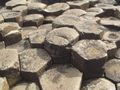
Hexamers are very common in nature. The bees use it in their hives. Giant's Causeway has volcanic basalt columns forming stepping stones that lead from the cliff foot and disappear under the sea. Most of the columns are hexagonal in form.


Then I remembered that some receptors have six proteine parts or helices, as acuaporines and gap junctions. Ordered in a ring structure like a hexamer. And the receptors are ordered in clusters, topologically arranged often. The neurons can even distinguish between left side of body and right side. The neurons/receptors are asymmetric.

About channels and their ligands(Ashcroft 2000)
Ligand-gated channels are generally named after the ligand that gates them. These may be extracellular, as in the case of the neurotransmitters acetylcholine and glycine, or intracellular such as cyclic AMP, Ca2+, and ATP. Binding of the ligand to one or more specific sites on the channel protein produce a conformational change that allosterically opens the ion pore, but sometimes close it.

At the resting potential of the cell, most voltage-gated channels are closed. When the membrane potential is changed, however. the channel undergoes a series of conformational changes that result in the opening of the channel pore. Also hyperpolarized gating exists.

The gating of almost all ion channels is subject to modulation by one or
more of a wide range of substances. These include monovalent and divalent cations (such as H- and Ca2-), metabolites such as ATP and MgADP, fatty acids, phosphorylation, GTP-binding proteins, and even gases (such as oxygen). The voltage dependence of activation is shifted to more negative membrane potentials in the presence of intracellular Ca2+. Sometimes the distinction between a channel modulator and the principal ligand seems merely semantic. Many hormones and neurotransmitters mediate their effects on ion channels indirectly by the activation of some second messenger system that modulates ion channel activity.

Chemotactic receptors
One of the best understood regions of cytoplasm is that associated with the cluster of chemotactic receptors in the plasma membrane of E. coli. Bacterial chemotaxis involves a phospho-relay system. This signaling complex transmits information from outside into the cell in the form of a phosphorylation signal that regulates flagellar rotation. The receptors are believed to exist in thermal equilibrium between two or more conformational states, and the output of the complex, usually measured in terms of phosphorylation levels is related to this equilibrium. The complex produces an amplified output proportional to the rate of change of attractant concentration.

The eukaryotic cell exhibits compartmentalization of functions to various membrane-bound organelles and to specific domains within each membrane.

An atomic level structure for a lattice of serine (Tsr) receptors are proposed. A unique feature of this model is that it creates a small compartment between the plasma membrane and an extended hexagonal lattice of the signaling proteins CheA and CheW. The compartment is not closed, and should be freely accessible to cytoplasmic proteins diffusing in from the lateral borders or through 10 nm diameter pores in the hexagonal lattice. This minute volume of bacterial cytoplasm will be highly enriched in two diffusible proteins = two enzymes that control the methylation of chemotactic receptors at sites in the middle of the receptor tails and have also been shown to carry binding sites for the C-termini of the receptor tails. It seems reasonable to suppose that they will accumulate in the small compartment because of this binding, and that their elevated concentrations in that privileged volume will facilitate the methylation and demethylation reactions.


The dynamics of the situation lead to the spontaneous emergence of order in the receptor lattice, such that receptors with 4 methyl groups (fully methylated) and receptors with 0 methyl groups (fully unmethylated) tend to lie next to each other in the array. The spontaneous emergence of order within a stochastically fluctuating field of allosteric proteins is an intriguing and potentially important phenomenon. By assigning different values of coupling energy to these different protein interactions and then examining the consequences for an array with the geometry of the receptor lattice the simulation model will be better.

This is fantastic. Sounds very TGD:ic in my ears. And reflexologic with all those geometric topologies.

Activity spread
The remarkable sensitivity and range of response of bacterial chemotaxis might depend on the clustering of chemotactic receptors on the surface of the bacterium. When a ligand bound to a receptor, the change in activity might propagate to neighbouring receptors in a cluster. Activity was spread by energetic exchanges between proteins in a two-dimensional lattice could be likened to the interactions of magnetic dipoles in a spin glass, resulting in digital receptors, also affected of the state of neighbours, "coupling energy".

Snapshot of an array of receptors showing a spread of activity. The receptors are portrayed as hexagons in an instantaneous snapshot showing their conformation as being either inactive (white) or active (orange).Bray et.al.1998.


Freeze fracture replica of two gap junctional plaques made up of a cluster of connexons (Ashcroft 2000),

Projected density map showing six-folded symmetry, rat Cx43 (Asgcroft 2000). See the neighbouring cells that are very sensitive to electric and magnetic changes. These cells are modulators (eg. their peptides).

Fluid mosaic membrane
It is assumed usually that the lipid membrane acts as an anchor for the proteins and has no independent function. However, membranes contain hundreds of different lipids (depicted in different colors in Fig.) which are different with respect to their physical properties (for example charge and size). One may wonder why nature has put so much effort into creating all these lipids if there is no functional purpose linked to their diversity. It is (for instance) known that bacteria change their lipid composition in response to changes in environmental conditions (e.g. different temperature of growth). Why is that, asks Heimburg?

Experiments and theoretical studies on model membranes, however, lead to the conclusion that the "fluid mosaic"-model requires major revision.


Modified version of the famous picture of the plasma membrane (Singer and Nicolson, 1972). Lipids (shown in different colors) are not uniformly distributed all over the membrane, but form domains of varying composition. Integral and peripheral proteins are depicted in green.

An important observation in a Monte-Carlo simulation (increasing temp.) is the formation of domains made of groups of gel or fluid lipids. Gel is in patches.
If proteins bind to a membrane with lipid domains, these proteins will be unevenly distributed on the membrane and the local protein concentration on the membrane surface may be quite variable. Membranes become soft and flexible in the transition regime.

Wave genome and phantom DNA?
Matti Pitkänen write: "Gariaev and collaborators have introduced the notion of wave genome requiring the coding of DNA sequences to temporal patterns of coherent em fields forming a bio-hologram representing geometric information about the organism. Code could mean that nucleotide is represented by a characteristic rotation angle for the polarization plane of linearly polarized laser radiation scattering from it. This kind rotation is known to be induced by chromosomes. Gariaev had photographs produced by the scattering of ordinary light on DNA, showing amazing "memory". In TGD framework these photographs could be interpreted as photographs of wormhole magnetic flux tubes containing dark matter."

The ability of this kind of light to induce gene expression in another organisms provided the modulated polarization pattern corresponds to an "address" characterizing the organism, and the formation of images of what is believed to be DNA sample itself and of the objects of environment by DNA sample in a cell irradiated by ordinary light in UV-IR range. This means that it works all ways, from within the cell reflekted out, or from outside reflected inside? A truly reflexologic picture.

What is even nastier is that in this way we can show how the DNA influences the cell membranes. And suddenly all those magnetic flux tubes and braidings makes sense. Even the magnetic body doesn't sound so peculiar. Biology is really quantum biology? As Matti said once "The guy was rightafter all?"

I can see how the chromosomes induces magnetic flux tubes, that are fastened to the receptors tails. And also to receptors on other membranes. By that neighbour-effect is then patches built on the membrane. That is phase-transitions, regulating the activity of the domain by inducing a gel. The signal reaching the membrane can be magnetic, electric or chemical (ionic) with hexamers as a vey important part. In fact life is about carbon, and carbon is about organic chemistry. In quantum physics there are the holy trinity, and a hierarchy of that forms hexamers.

Perhaps Matti he himself will explain these things in more detail to us when he has time for it. We are waiting for that impatiently.

What has all this in common with the Saturn pole? It is ice, or water. More about that later.

References:

Acshcroft 2000: Ion channels and disease.

Bray et.al. 1998, www.pdn.cam.ac.uk/groups/comp-cell/Papers/Bray98a.pdf

Crystal structure of a molecular hexagon composed of hexagonal aromatic rings reported by Müllen and coworkers in Chem. Eur. J., 2000, 1834-1839.
http://en.wikipedia.org/wiki/Coronene

Cassini Images Bizarre Hexagon on Saturn 03.27.07, http://www.nasa.gov/mission_pages/cassini/media/cassini-20070327.html

Heimburg,T. 1998?: AG 012 - Membrane Thermodynamics. The fluid-mosaic model. Fluctuations and Domains: Thermodynamic properties and heterogeneity of membrane assemblies. http://membranes.nbi.dk/presentation-english/News_engl.html

Maddock, J. R., & Shapiro, L. (1993). Polar location of the chemoreceptor complex in the Escherichia coli cell. Science 259, 1717-1723. http://www.sciencemag.org/cgi/content/abstract/259/5102/1717

Pitkänen, M. 2008: Has dark matter at the magnetic flux tubes been photographed? http://matpitka.blogspot.com/2008/05/has-dark-matter-at-magnetic-flux-tubes.html.

Receptor Clusters, 2006. Bray Group: Computer Models of Bacterial chemotaxis, Physiology, Development and Neuroscience, University of Cambridge, http://www.pdn.cam.ac.uk/groups/comp-cell/Cluster.html

fredag 1 maj 2009

Singing, screaming and shouting cells

Communication is complicated. So also for the various cells in our body. How could such completely different cells as neurons and let us say bonecells, or highly undifferentiated mesenchymatic cells "talk" to each other? What kind of language can they possibly use? At the Center for Models of Life they talk about decision taking.

One guru in this scientific area is Candice Pert. Her "Molecules of emotion" had a profound impact on me. She described this communication as a singing process, and the main media are peptides. She describes the cellmembrane as a veritable forest. The cell is communicating using frequensies, and a healthy cell use harmonious frequencies. What is harmony in this sense? Of course fractality. Looks like our body is fractal in every possible way? Structurally and functionally.

We are used to think of this "talk" in terms of nerves and hormones. Nerves "talk" by a chemical impulse, have we learned. But how this impulse can "tell" the information to its surroundings is still a mystery. In an impulse the only thing that is propagated is "noise". The neuron itself doesn´t change (but the synaptic/dendritic ends do change = LTP), or let us say the cellmembrane doesn´t change. The wave carry the information only, as a disturbance or noise. How can this information be encoded? Different frequencies? Perhaps we can compare to a computer lightfiber transfer?

The nerve pulse is frequential, not chemical. But how on earth do the synaptic endings know what kind of neurotransmitters to release, and how much, and when? Like all other cells the neurones have just one nucleus for proteine synthesis, or for making copies of DNA in form of single-helical mRNA. Just how is this mRNA transported to the synaptic bouton, which can be a very long distance, and exactly what regulate the proteine synthesis? Today we have very few answers to these questions.

One kind of answer is that the nerve impulse is in reality sound. This is explained by the alternative soliton-impulse theory proposed by Thomas Heimburg and collaborators at Niels Bohr Institute in Copenhagen. A density-pulse of different phases in the cellmembrane. During the pulse the membrane"halves" (lipidsheats) widens a little, but no increased heat is observed. On the countrary the membrane is cooled. Something is not what it should be here. Heimburg suggest we have ended up with the wrong theory for the nerve pulse. The impulse is sound, and a quantal one, using Josephson-currents and phase-transitions. The membrane biophysics group writes: "During the action potential in nerves one finds a reversible heat change and mechanical changes. This indicates that the physics underlying the nerve pulse relies on reversible physical processes. We recently explored the possibility that the nerve pulse is in fact a propagating density pulse in the nerve membrane (soliton). Such pulses can propagate close to chain melting transitions in cylindrical membranes. These pulses show many properties that have been experimentally measured, like reversible heat production and thickness changes associated to the nerve pulse. This theory yields a surprisingly simple explanation for anesthesia. It acts as a feezing point depressant for biomembrane transitions and renders nerve pulse generation more difficult."

This however explaines no more than the old theory of Hodgkin & Huxley about the functions of synaptic boutons. On the countrary we also gets the mysteries with proteines in the cell membrane, and the real functionings of receptors in the membrane. With the solitonic explanation there is no need for a lot of energy. What is the energy used for? Why so much mitochondrions in axones? The role of free radicals? How do the myelin work? What is the role of membrane receptors? How do they transfer their information? And how do the cells discriminate among all songs, shoutings and screams from all the different boutons (synchronization and organization)? This all points to a very strong electromagnetic function among receptores, neurotransmitters and other signal transduction matters as protons, electrons and calciumions.

Temperature, pH, and mechanical force among other factors interfere with the solitone. Topology is a word of honor. The relationship between the regulatory design and the functionality of molecular networks is a key issue in biology, says Axelsen. Rewiring of regulatory links plays a bigger role than genes. Many feed-forward loops works in transcription networks.

Epigenetics
Cells carry information handed down from their ancestors and are able to pass on information to their descendants. In many cases this ``memory" is epigenetic -- not stored in the DNA sequence -- allowing cells with identical DNA to maintain distinct properties. Epigenetic cell memory implies alternative states that are stable over time and are inherited through cell division. Mechanism for epigenetic cell memory invokes positive feedback loops, either on genes with mutual repression or, for eucaryotes, through recruited modifications of DNA bound nucleosomes. Thus, positive-feedback loops in nucleosome modification can, in theory, provide not only a mechanism for long-term epigenetic memory (Dodd), but also a powerful system for controlling the way in which a promoter gene responds to and integrates multiple signals.

Triggerpoints
Skeletal myofibrils spontaneously oscillate when free Ca2+ is low (in resting state), was reported by Ishiwata 1996.. Although this oscillation phenomenon called SPOC is apparently simple, the molecular mechanism seems to be complex. It is regulated by the mechanical strain imposed on actin and myosin; the enzymatic activity (ATPase) of actomyosin complex and the mechanical event (contraction) are thus coupled to each other.

Ysuda et.al. 1996. The change of sarcomere lengths in glycerinated skeletal myofibrils oscillated spontaneously with a peak-to-peak amplitude of about 0.5 microns under isotonic conditions in which the external loads were maintained constant. The shortening and yielding of sarcomeres occurred in concert, in contrast to the previously reported conditions (isomeric or auxotonic) under which the myofibrillar tension (connectin, an elastic proteine)is allowed to oscillate. This synchronous SPOC appears to be at a higher level of synchrony than in the organized state of SPOC previously observed under auxotonic conditions. The period of sarcomere length oscillation did not largely depend on external load. The synchronous SPOC implies that there is a mechanism for transmitting information between sarcomeres such that the state of activation of sarcomeres is affected by the state of adjacent sarcomeres. The change of myofibrillar tension is not responsible for the SPOC of each sarcomere but that it affects the level of synchrony of sarcomere oscillations. Declined synchrony is the result of increasing tension. A certain degree of strain is demanded, though.

Mechanical strain is also sometimes behind triggerpoints. Triggerpoints are not the same thing as acupuncture points. If we look at the points histochemically we can see this clearly. Triggerpoints are scars in myofibrils, whereas acupoints are distinct anatomical structures.

Muscles contract when, in response to a motor nerve signal, calcium is released from storage in the sarcoplasmic reticulum. In the resting muscle, ATP is bound to myosin in a high energy configuration. The myosin cannot however do anything until calcium is released from the sarcoplsamic reticulum. The calcium switch is turned off. Muscle in Rigor Mortis - the actin-myosin unit is locked together and cannot release: The calcium switch has turned on, and has enabled the high energy myosin-ATP to "bind and bend" to the actin with the release of low energy ADP and Phosphate. However that is as far as it goes: there is no more energy currency (ATP) in the muscle cell to drive the myosin cycle of "release and straighten, bind and bend", and the myosin remains stuck fast to the actin molecule. In active muscle - both calcium and ATP are present, and the myosin cycle of "Release and straighten, bind and bend" is in full flight. The myosin is "walking up the actin". Normal muscle has sufficient ATP in the cell and sufficient integrity in its sarcoplasmic reticulum to quickly re-absorb the calcium and turn off the contraction. However, a trigger point zone in a muscle cannot re-absorb the calcium and turn the contraction off: The contraction continues until tension pulls hard enough against the myosin "leg" to stop it at the bending phase of its cycle. Little wonder there is intense activity with build up of lactic acid and molecular damage. Necroses may be the result.

A Trigger Point in a Muscle, severe damage at the Sub-cellular and molecular levels has been noted. Microsocpic examination of trigger point sarcomeres reveals signs of damage to actin fibers. Triggerpoints are identified by what is called Travelli's twitch, a small reflexive contraction (motoric) of the muscle. The autonomous nervous system is one part of the trigger. EMG-response is much higher. Triggerpoints are not restricted to the muscles, but can be found in all kind of connective tissues, like scars, fascias, tendons, periosteum, joint capsules... Connective tissue is alpha-helical too, like actin.

Muscle force generation has been interpreted traditionally on the basis of the kinetics of crossbridge cycling, i.e. binding of myosin heads to actin and consecutive force generating conformational change of the head. However, several studies have shown that re-distribution of internal strain within myofibrils and muscle fibres may be a key player, particularly, during stretch or relaxation so that force kinetics parameters are strongly affected by sarcomere dynamics, says Telley IA & Denoth J in a rewiev-article 2007.

If we only knew why the calcium switch fails to turn off! Actin seems to be the important part (I-band). This amplification of coordinated cross-bridge binding and cycling indicates a mechanism of cooperativity that depends on sarcomere lattice geometry, specifically the ratio and arrangement of myofilaments. Topology is again very important.

Calciumwaves
The most important ion when talking about communication is calcium.
A large number of ion channels, enzymes, pumps and binding proteins participate in the generation of intracellular Ca2+ signals and their decoding. Ca2+ signalling takes place in the form of oscillations, waves and sparks. Such Ca2+ signals occur in almost all cells and regulate diverse cell functions. Perturbation of Ca2+ signalling leads to disease. Drugs that act on Ca2+-signalling are commonly used in treatment of several diseases.

This Ca-wave also mediate an metabolic wave of insuline and glucose-metabolism (for ATP/GTP?). Other ions as Na and K are also oscillating.

It acts as second messenger in the dendritic nerve loop. This is how the nerve pulse message is propagated? The most wellknown Ca2+-canals is activated of depolarization.

A revolution has happened.
Glial cells are very active parts of the nerve-pulse. Astrocytes organize synapses. They control neurotransmitter release and free calcium in the synaptic cleft through calciumwaves of their own. And neurotransmitters evoke calcium waves. "The existence of bidirectional signaling between astrocytes and neurons has revealed an important active role of astrocytes in the physiology of the nervous system. As a consequence, there is a new concept of the synaptic physiology-"the tripartite synapse", where astrocytes exchange information with the pre- and postsynaptic elements and participate as dynamic regulatory elements in neurotransmission. The control of the Ca2+ excitability in astrocytes is a key element in this loop of information exchange. The ability of astrocytes to respond to neuronal activity and discriminate between the activity of different synapses, the modulation of the astrocytic cellular excitability by the synaptic activity, and the expression of cellular intrinsic properties indicate that astrocytes are endowed with cellular computational characteristics that process synaptic information. Therefore, we propose that astrocytes can be considered as cellular elements involved in the information processing by the nervous system." Says Perea & Araque 2005.

We now get a picture where the actual decision making is done not by neurones but by glial cells. Neurones are simply a tool for glial cells? There is something else that gives orders to our nerves.

Nanomechanical motion
Pelling et.al. demonstrated that the cell wall of yest, Saccharomyces cerevisiae, exhibits local temperature-dependent nanomechanical motion at characteristic frequencies (0.8 to 1.6 kHz with amplitudes of ~3 nm), and a metabolic inhibitor causes the periodic motion to cease. An calculated activation energy of 58 kJ/mol is consistent with the cell's metabolism involving molecular motors such as kinesin, dynein, and myosin. The magnitude of the forces observed (~10 nN) suggests concerted nanomechanical activity. These examples of biological processes involving concerted motor protein action lend strong support to our conclusion that a metabolically driven nanomechanical process occurs at the yeast cell wall. This process cannot be observed by traditional cytological methods and occurs in cells in their natural state. The observed motion may be part of a communication pathway or pumping mechanism by which the yeast cell supplements the passive diffusion of nutrients and/or drives transport of chemicals across the cell wall. The current experiments were performed on yeast cells because they have a stiff cell wall. Extension of this experiment to mammalian cells will require the use of specially fabricated cantilevers with small spring constants comparable to the spring constant of the mammalian cell membrane (~0.002 N/m).

Microtubules.
In close connection with the membrane are microtubules (MTs). Also other kinds of tubuli are there. Cytoskeletal elements interact extensively and intimately with cellular membranes. Microtubules are components of the cytoskeleton, the network of proteinaceous fibers that endows the cell with structural integrity, motile properties, and internal organization.

MTs play a particularly important role in cell organization: they pull the chromosomes apart at mitosis, act as a ‘railroad system’ for intracellular transport, and define the localization and structure of internal membrane systems. Two characteristics of MTs are particularly significant for these functions. First, MT nucleation is regulated, and the limitation of nucleation to the centrosome endows most cell types with a radial organization. Second, and perhaps more importantly, the MT cytoskeleton is dynamic: individual MTs in the same cell (or same test tube) constantly change in length, either growing or shrinking with random transitions between these phases. This counterintuitive behavior is termed dynamic instability. I would say cellular "brain". First, dynamic instability is a mechanism for exploring cellular space, bringing MT railroads into contact with poorly diffusible cargo, such as chromosomes for subsequent transport, Second, this turnover ensures rapid response of the cytoskeleton to internal and external signals. Selective stabilization of dynamic MTs probably plays a key role in morphogenesis and appears to play a central role in the selforganizing properties of the mitotic spindle. Briefly, tubulin subunits (which are obligate dimers of the polypeptides alpha- and beta-tubulin) bind GTP. Upon polymerization, this GTP is hydrolyzed to GDP, but only after a short delay. This delay is thought to result in a ‘GTP cap’,which predisposes the MT to continued growth.

In addition to movement generated by the dynamic instability of the microtubule itself, the fibers are substrates along which motor proteins can move. The major microtubule motor proteins are kinesin, which generally moves towards the (+) end of the microtubule, and dynein, which generally moves towards the (−) end.

Microfilament.
Actin is the monomeric subunit of two types of filaments in cells: microfilaments, one of the three major components of the cytoskeleton, and thin filaments, part of the contractile apparatus in muscle cells. Thus, actin participates in many important cellular processes including muscle contraction, cell motility, cell division and cytokinesis, vesicle and organelle movement, cell signaling, and the establishment and maintenance of cell junctions and cell shape. Many of these processes are mediated by extensive and intimate interactions of actin with cellular membranes.

Around 6 nm in diameter, actin filament type is composed of two intertwined actin chains. Microfilaments are most concentrated just beneath the cell membrane, and are responsible for resisting tension and maintaining cellular shape, forming cytoplasmatic protuberances.

Endolysosomal tubules in dendritic immunocells are dynamic and display saltatory, bidirectional movement. These tubules have a functional dependence on microtubule tracts and can use polymerizing microtubules.

Nanotubules are discussed earlier in this blog.


Model of endolysosomal tubules and interdependence with microtubules.
Operating speeds of 0.2 to 8 µm s–1 for the myosins and 0.02 to 7 µm s–1 for the microtubule proteins kinesin and dynein have been reported, and many processes taking place inside the cell are mediated by these proteins, with operating speeds 1 to 2 orders of magnitude faster than the individual motor proteins. The force being generated at the cell wall can be determined to a value of 0.2 nN. We can exclude that a single motor protein is driving the observed nanomechanical motion, because the forces observed at the cell wall (~10 nN) are far too large in magnitude. Large-scale forces are generated in yeast cells through the action of many proteins working in a concerted and cooperative manner?

The best-studied molecular motors, myosin from skeletal muscle and conventional kinesin from brain, are remarkably similar in structure, yet have very different functions. These differences can be understood in terms of the 'duty ratio', the fraction of the time that a motor is attached to its filament. Differences in duty ratio can explain the diversity of structures, speeds and oligomerization states of members of the large kinesin, myosin and dynein families of motors. Howard J. 1997.

Is the subjective I a timelag?
Microtubules are the "brain" of the cell. They are doing the computating and desicion taking. And then the cells inform the rest of the body about their desicions through Em-waves, or sound, light etc. The result is then coordinated in waves like EMG, EEG and so on. But there is (always) a "time lag phase" in the pulse, like that "duty ratio" above. Also Benjamin Libet found this delay (350 - 500 msec.). He demonstrated that the readiness potential precedes the time at which participants consciously decide to perform an intentional motor act. Neural activity was preceding the motor response, similar to Libet's experiments; this means the sensory world is experienced delayed with respect to real time. However, this activity was already present prior to stimulus presentation, and thus before participants could decide which button to press. New research has identified networks of brain areas that underlie voluntary action. These areas generate information for forthcoming actions, and also cause the distinctive conscious experience of intending to act and then controlling one's own actions. Automatic motor activation (reflexes) forms an intrinsic part of all behavior, rather than being categorically different from voluntary actions. A crucial issue is how such automatic mechanisms are controlled so that the most appropriate responses are made and unwanted responses inhibited.

If we see the subjective I (and perhaps the consciousness in biological way) as a disturbance of the cosmic Em-waves, life itself is indeed very much the same as these oscillations. Incoming oscillation in kHz for Ca, or even in GHz for 1 - 10 mm-waves (the extremely-high-frequency (EHF) band: f = 300 - microwave - to 30 GHz - IR - optical wave). This is then reduced in DNA to UV-band-frequency. And from DNA further reduced in EMG; the bigger the disturbance the bigger value. Eye has one of the biggest frequencies, together with the hearth. They are doing very hard work.
Further reduction of the disturbance is seen in EEG, with the highest value, 40 Hz, for incoming disturbances. This is then reduced to alpha-waves about 10 Hz. This can be seen as the ultimate brainwave, and is only slightly above Schumann resonance waves of Earth. Indeed there are also lower frequencies. Josephson oscillate at somewhat above 3 Hz.

What is characteristic for these waves? Of course their information-content, or their synchronic and ordered behaviour, their harmony. If these waves are interrupted in the brainstem they are loosing this ordered behaviour and is becoming desynchronized or global. Then the patient is in coma, but not necessarily without consciousness.

Desynchronized waves are screaming?


Rabbit horizontal cell network revealed by dye injections. The dye spreads via the gap junctions linking the horizontal cells to reveal the centrally injected cell and hundreds of neighbouring cells.

Plasmatic network and hemichannels syncytia.
Coupling seems essential to maintain this oscillatory behaviour, as isolated cells are unable to oscillate. Coupling coefficients and coupling conductances changes dynamically and in phase with the membrane potential oscillations, pointing to an active modulation of the gap junctions. Nanotubes and tunnels are connecting different cells into a cell - cell network through gap junctions. The role of the junctional couplings is to synchronize the oscillations, interfere with the ionic permeabilty and intracellular messages. Emerging evidence suggests that gap junction hemichannels can act as stand-alone functional channels in astrocytes. They can also mediate robust efflux of glutamate, aspartate, and other messages. Also viable proteines can transinfect cells, as in the Rhett syndrome. Chaperones, kinesines, TNFs, cytokines, ILs, PGLs and many, many more signal substances, voltage and metabolites, even oxygen and free radicals, work in intimate connection to this network. It is really question of sick or health states.

'When a "tape" of mRNA passes through the "playing head" of a ribosome, the "notes" produced are amino acids and the pieces of music they make up are proteins.' You get "DNA-songs".

Referenses:
Axelsen, Jacob Bock et.al. 2008: One hub-one process: a tool based view on regulatory network topology. BMC Systems Biology 2008, 2:25. http://www.biomedcentral.com/1752-0509/2/25

Dodd IB, Micheelsen MA, Sneppen K, Thon G (2007) Theoretical analysis of epigenetic cell memory by nucleosome modification. Cell 129: 813–822 http://www.nature.com/msb/journal/v4/n1/full/msb200821.html#B7

Libet B, Gleason CA, Wright EW, Pearl DK. 1983: Time of conscious intention to act in relation to onset of cerebral activity (readiness-potential). The unconscious initiation of a freely voluntary act. Brain. 1983 Sep;106 (Pt 3):623-42.

Nick, P. 1998: Signals, Motors, Morphogenesis — the Cytoskeleton in Plant Development. Institut für Biologie II, Freiburg, Germany
Received: Sept 25, 1998; http://www.rz.uni-karlsruhe.de/~db45/Publikationen/Nick%201999.pdf

Milo, R. 2002: Network Motifs: Simple Building Blocks of Complex Networks. Science 25 October 2002: Vol. 298. no. 5594, pp. 824 - 827. http://www.sciencemag.org/cgi/content/full/298/5594/824

Sneppen, Kim, Mille A Micheelsen & Ian B Dodd (2008).Ultrasensitive gene regulation by positive feedback loops in nucleosome modification. Molecular Systems Biology 4. http://www.nature.com/msb/journal/v4/n1/full/msb200821.html

Pelling, Andrew E., Sadaf Sehati, Edith B. Gralla, Joan S. Valentine, James K. Gimzewski 2004: Local Nanomechanical Motion of the Cell Wall of Saccharomyces cerevisiae. Science 20 August 2004: Vol. 305. no. 5687, pp. 1147 - 1150. http://www.sciencemag.org/cgi/content/full/305/5687/1147

Telley IA, Denoth J. 2007: Sarcomere dynamics during muscular contraction and their implications to muscle function. J Muscle Res Cell Motil. 2007;28(1):89-104.

Travell & Simons' Myofascial Pain and Dysfunction: The Trigger Point Manual (2-Volume Set.)David G. Simons (Author), Janet G. Travell (Author), Lois S. Simons (Author), Barbara D. Cummings (Author).

Yasuda, K, Y Shindo, and S Ishiwata 1996: Synchronous behavior of spontaneous oscillations of sarcomeres in skeletal myofibrils under isotonic conditions. Biophys J. 1996 April; 70(4): 1823–1829. http://www.pubmedcentral.nih.gov/articlerender.fcgi?tool=pubmed&pubmedid=8785342

söndag 22 mars 2009

Genkoden

biology200.gsu.edu/.../4564%20'04/lecture12.html Största delen av genomet består av annat än gener.

Det är ett allmänt känt mysterium att vi har för litet gener. Vartefter genkartan blivit känd har antalet blivit mindre och mindre, från c. 100000 till i dag litet över 20000.

Det som skiljer en mänska från en schimpans är också otroligt litet, bara några baspar, 0,6 %. Tidigare trodde man att skillnaden, c. 1,5% av generna, skulle kunna förklara skillnaden, ex. med sk. "nyckelgener". Vartefter schimpansens DNA kartlagts har man kommit underfund med att så inte är fallet. Utvecklingen från schimpans till mänska har krävt också annat än genmutationer.

Gener är starkt överreklamerade. Mänskans DNA är kartlagt, har man trumpeterat ut. Man funderar tom. på att patentera gener. Allt detta är vilseledande tomt prat, kanhända för att få forskningspengar. I dagens läge är gener rätt säkra forskningsobjekt, just genom lättheten att få finansiering.

DNA består mest av - nonsens. Bara 1,2% av DNA:t är gener, extroner. Resten är "skräp", vilket är en felaktig term, för skräpet, intronerna, är den del av genomet som är den viktiga biten. Hela 98,8 % av DNA:t är okänt. Det kodar annat än gener, ex. olika RNA. Också gener kodar RNA för den delen, sk. messengerRNA. Men RNAs produkt är så mycket mera än proteiner. Small RNA är ett ex. De ger en sorts primitivt immunsystem, som man tror fanns innan det immunsystem vi har i dag fanns. Den här sk. interferensen upptäckte man av en slump när man ville få fram extra mörkblåa petunior. De blev till allas förvåning vita, och så fick man upp spåret till detta stora mysterium. En google-sökning i dag på siRNA ger miljoner träffar. Det är en forskning i stark expansion.

Fenotypen är inte alltid beroende av genomet. Kallas non-Mendelisk nedärvning. I det här fallet påverkas slutresultatet (fenotypen) av ett intensivt informationsutbyte mellan kromosom och plastid, ex klorofyll, mitokondrie.
www.biologie.uni-hamburg.de/b-online/e10/10e.htm

Evolutionen tar inte bort någon viktig funktion i första taget, utan funktionerna läggs till. Det kan inte heller vara på något annat sätt, för om en funktion tas bort innan en ny funktion prövats fram genom försök och misstag, kanske den nya funktionen inte visar sig vara livsduglig, och då slutar det illa. Det är inte bara RNA som adderas, utan det gäller alla funktioner. Nervsystem (DC-elektriskt system, nanotuber, meridianer), hormoner (autokrina hormon), signalämnen (eikosanoider) mm. har alla ett modernt system och ett eller flera primitiva system (de inom parentes) som fungerar sida vid sida. Ofta har det nyutvecklade systemet fungerat nästan parallellt med det gamla som därigenom undgått upptäckt, vilket är fallet med nervsystemet. Jämsides med vårt digitala nervsystem (on-off-system) finns det ett analogt likströmssystem. Och dessutom har vi ett "våt-elsystem" i de sk. meridianerna, som kan öppnas eller stängas i sin helhet via piezoelektriska signaler. Gap junctions m.fl. desmosomer är en led i detta system. Här syns igen den fraktala egenskapen i mänskokroppen? Man kan kanske jämföra med en mobiltelefon där över 10 tunna kretskort läggs ovanpå varann. Detta syns inte i funktionen alls.

Skillnaden mellan schimpans och mänska belyser detta på ett bra sätt. Skillnaden finns i intronerna, eller i hur generna används. Mänskans DNA är mera flexibelt än schimpansens DNA. Eller eg. mänskan har ett mera anpassningsbart sätt att använda generna. Intronerna bestämmer hur generna används. Samma gen kan ingå i flera olika protein, och därför kan genprodukten mångdubblas i det sk. proteonomet. Och ännu mera i metabonomet.

Genkoden är inte alls enkel. Finns det flera olika genkoder? Den nuvarande genkoden med tripletter ger 64 möjligheter, men bara 20 aminosyror + start. 3 st möjligheter saknas? I dag finns misstanken om att alla aminosyror inte är likvärdiga, trots att de räknas till samma aminosyra. Den sista koden skiljer? En annan typ av kod kan vara ex. purin-pyrimidin (energikällor). Metylerade (platsen viktig) puriner bryts ner, hos pyrimidiner bryts ringen upp, reduceras av NADPH. Eller topoisomeras-enzymet som får DNA-topologin att ändras? Tyrosin katalyserar omformningen, "klippningen" och avkodningen? Stressen ökar på DNA, konfigurationen ändrar. Nukleosomer med histoner är naturens svar. De låser DNA:t. Allt detta är mycket intressant och har fått ett eget namn; epigenetik. Eller hur generna aktiveras eller inaktiveras. Det är inte generna som är de intressanta delarna, utan det som styr genernas uttryck.

Ser man på intronernas sekvenser blir man inte klok. En upprepning ofta av samma kodord. De liknar mycket telomerernas koder, och telomererna vet vi är ytterst viktiga för kromosomens avkodning vid replikationen. Avkodningen vid syntes av genprodukt styrs enligt samma typ av mekanism, en elektromagnetisk våg? Telepatiska gener? Identiska basmönster förenar?

Intronerna, i dag kallat tilke-DNA, utfyllnads-DNA, kan härstamma från virus. Man har funderat om virus på detta sätt kan gömma sig, för att en dag igen vakna till liv. Gamla, utdöda farsoter kanske en dag oväntat blossar upp på nytt? En stor del av intronerna misstänker man består just av sådant som förändrats så att det inte mera fungerar, ex doftsinnet. Hos däggdjuren fungerar c. 1000 gener, hos schimpanser fungerar bara 65 % mera, hos mänska 40%. Resten är icke funktionsdugliga minnen ur det förflutna? Men kan dessa minnen ibland aktiveras? Växterna kanske har svaret. Samma gener styr utvecklingen av hjärtblad och blomma. För att inte tala om insekternas metamorfos. Fraktala funktioner igen?

Sjukdomar som mänskan, och dess förfäder haft, kan berätta mycket om intronerna och genregleringen. En utdöd sjukdom kanske bekämpades med en viss molekyl. Schimpanserna har ex. en gen som bildar sialinsyra, en antiviral komponent. Mänskan saknar denna gen. Mänskan får mera virussjukdomar, ex. AIDS? Sjukdom befrämjar mutationer, vilket man sett gälla också hos bakterier. De har satt detta i system, så att då någon fara hotar startar en formidabel "mutationsexplosion". Alltid är det någon av de nya varianterna som fungerar.

Schizofreni är en sjukdom som allmänt anses vara en orsak till att arten mänskan föddes. Genier och de schizofrena har något gemensamt.

Också matvanorna syns i intronerna. Maten kan ha gjort så att hjärnan började växa någon gång för 70000 milj.år sedan. Omega 3 havsoljor är en sådan faktor. Därför är omega 3 så hälsosam i dag, och vi äter för litet havsmat, eftersom våra kostrekommendationer är felaktiga. De rekommenderar på tok för mycket kolhydrater. Inte underligt alls att en av faktorerna bakom schizofreni misstänks vara brist på omega 3? Omega 3 från växtriket är inte alls samma sak. De är för långa kedjor. De måste bearbetas extra, och det kräver energi. Nyttan blir då inte samma sak. Och dessutom har oljor från ex.lin och ryps sina egna negativa biverkningar, ex. i fråga om sköldkörtelfunktionen.

Alla däggdjur har FOXP2-genen men det är bara mänskan som kan tala. Samma gen har flera olika funktioner. Hos mänskan har den muterat så att basparen ligger annorlunda. Men skillnaden mänska - schimpans är bara två mutationer som skett för kanske 200000 år sedan. De har gett mänskan egenskaper som hjälpt fram talet?

Mikrocephalia- genen bestämmer hjärnans storlek. Fel på den ger liten hjärna och dvärgväxt. En annan gen bidrar också, MYH-16, vilken producerar proteiner som reglerar käkmuskulaturen. Hos mänskan fungerar inte dessa gener. Köttätandet har bidragit, då de inte längre behövdes, och så muterades de och mutationen korrigerades inte. Hjärnan fick mera rum att växa. Den andliga tillväxten kunde börja. Men generna i sig kan inte ensamma förklara detta.

En stor hjärna kräver mycket energi. Dessa gener måste då omformas. Under utvecklingens gång har de gener som styr hjärnaktiviteten fyrdubblat sin aktivitet. DNA är inte så viktigt? Det viktiga är hur det används? Cis-reglerelement styr? Mänskan har stor varians i sina cis-element. Mänskans genom är mera kreativt och har större möjlighetspotential? Frontalloben och neocortex uttrycker en fraktal funktion?

De funktionella begränsningarna mot olika proteiner har blivit slappare hos mänskan. Vi kan tillåta oss en större variation än schimpansen? Vi har det bättre ställt. Nukleotidsekvenser borde ställas mot fysiologi och biokemi, enl. Satta.

Mänskans nervsystem innehåller c. 100 miljarder celler av 10000 olika typer, men det finns bara ett par tusen reglerande gener. Hur avgörs ödet för en enskild cell? Processen sker i en regleringskedja genom samverkan av 4-7 olika gener. Det kan liknas vid en kryddhylla, där varje krydda har sin egen smak, och blandningar ger sina egna sensationer. Rönen gäller två typer av nervceller hos bananflugor, men angående funktionen skiljer vi oss inte så mycket från bananflugorna.

En av de stora skillnaderna är mänskans behov av att förstå sig själv och sin omgivning. Tolkningarna, de sk. qualias, fenomenen, är viktiga, och här använder vi spegelneuroner som hjälp. Spegelneuroner får sin utformning under barnaåren. Autister har dåligt fungerande spegelneuroner. Det är en fråga om medvetande. Uppfattningsförmågan eller perceptionen, som för det mesta sker i det undermedvetna? Medvetenheten och medvetandet är två olika saker. Signalintensitet som en storskalig integration, eller timing (impulsfrekvens, amplitudfrekvens?) eller något annat? Kanske magnetism? Funktionella cortikala områden, sk. Zeki-noder? Kandidaterna är många.

Referenser:
Angstadt, Carol, 1997: Purine and pyrimidine metabolism. NetBiochem. http//library.med.utah.edu/NetBiochem/pupyr/

Forbes, Ken, 2005: You Descend from Banana or Bonobo? GENEALOGY-DNA-L Archives 2005-01 > 1106711681

Kornyshev, Alexei, 2008: 'Telepathic' genes recognize similarities in each other. BJS. http://www.imperial.ac.uk

Priemé, Anders2005: Se tärkeä ero. Tieteen kuvalehti 2005(2):36-41.

Rees, Geraint et.al. NEURAL CORRELATES OF CONSCIOUSNESS IN HUMANS. www.nature.com/reviews/neuro Nature review, APRIL 2002, VOL 3: 261- 270.

Satta, Yoko, 2001: Comparison of DNA and protein polymorphisms between humans and chimpanzees. Genes & Genetic Systems Vol. 76 (2001) , No. 3 p.159-168

Thor, Stefan 2007: Genkod för nervceller knäckt? Forskning.se, 22.2.2007. Pressmeddelande från Linköpings universitet. PLoS Biology febr. 2007. Magnus Baumgardt et.al. Specification of neuronal identities by feedforward combinatorial coding.